Copyright
©The Author(s) 2022.
World J Clin Cases. Jul 26, 2022; 10(21): 7459-7466
Published online Jul 26, 2022. doi: 10.12998/wjcc.v10.i21.7459
Published online Jul 26, 2022. doi: 10.12998/wjcc.v10.i21.7459
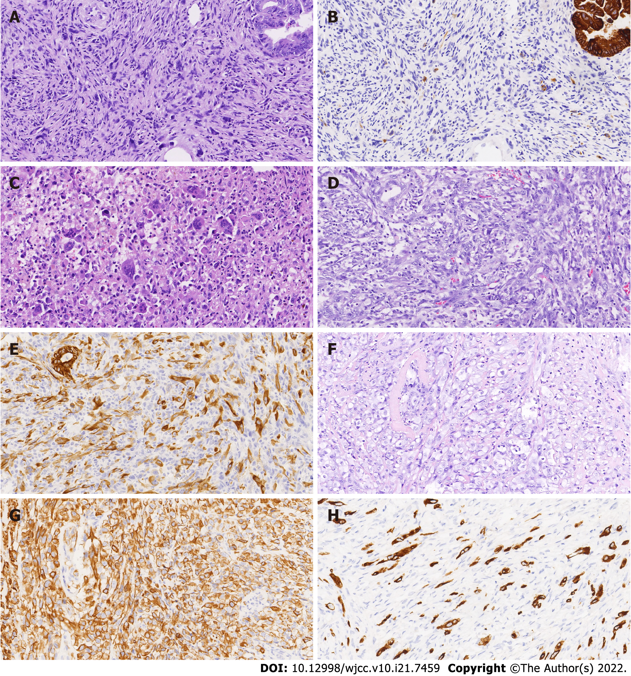
Figure 1 Microscopic morphology and immunohistochemical results of mural nodules in three cases of anaplastic carcinoma.
A: Sarcoma-like mural nodules (SLMNs) of case 1 (20 ×); B: Expression of AE1/AE3 in SLMN of case 1 (20 ×); C: Osteoclast-like multinucleated giant cells in SLMNs of case 1 (20 ×); D: Mural nodule of anaplastic carcinoma in case 1 (20 ×); E: Expression of AE1/AE3 in anaplastic carcinoma in case 1 (20 ×); F: Mural nodule of anaplastic carcinoma in case 2 (20 ×); G: Expression of AE1/AE3 in anaplastic carcinoma in case 1 (20 ×); H: Expression of AE1/AE3 in anaplastic carcinoma in case 3 (20 ×).
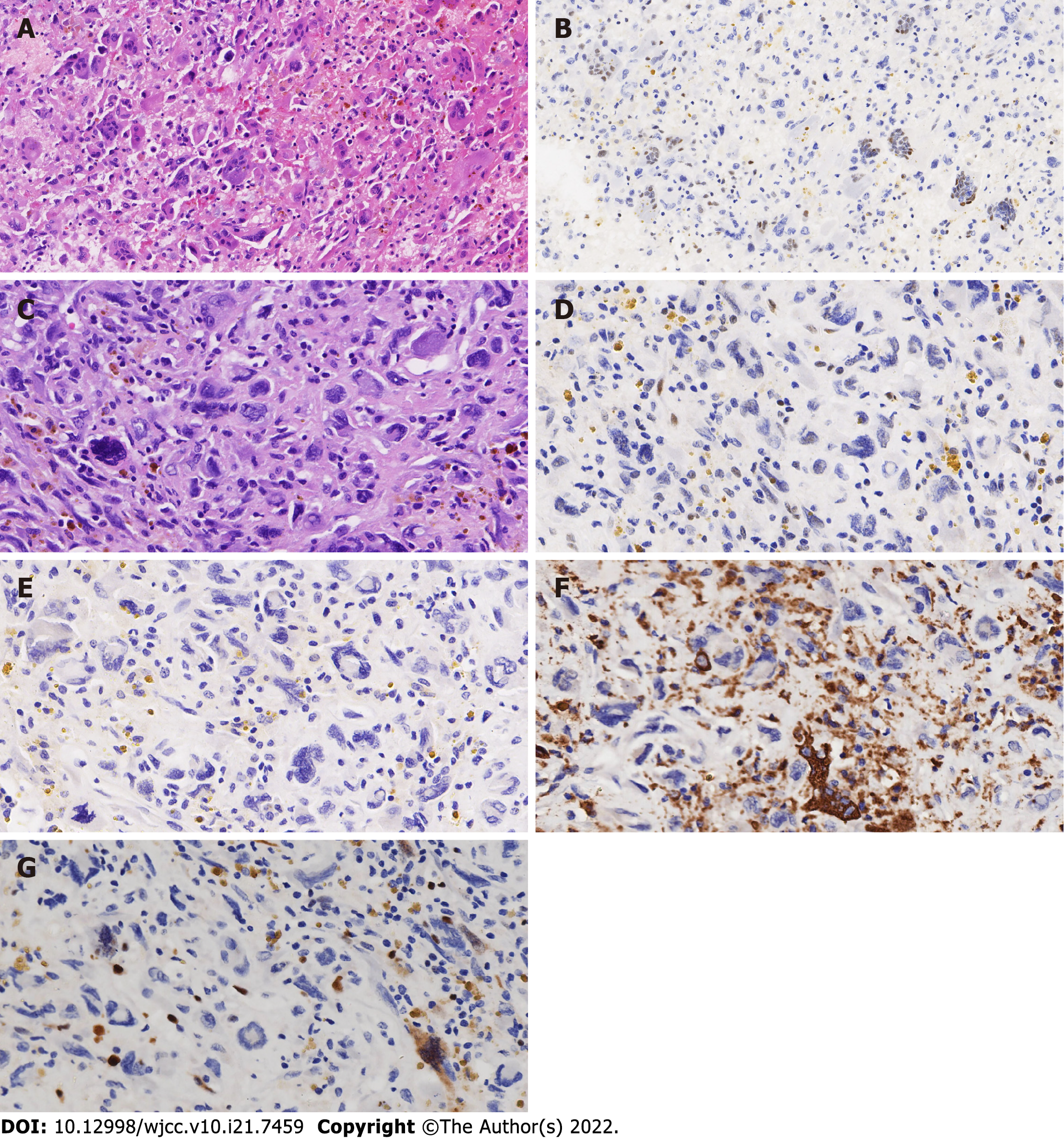
Figure 2 Two types of giant cells in the sarcoma-like mural nodules of case 1.
A: Osteoclast-like multinucleated giant cells (20 ×); B: Wild-type expression of TP53 (20 ×); C: Another kind of giant cells (40 ×); D: Expression of TP53 in another kind of giant cells (40 ×); E: Expression of AE1/AE3 (40 ×); F: Expression of CD68 (40 ×); G: Expression of Ki-67 (40 ×).
- Citation: Wang XJ, Wang CY, Xi YF, Bu P, Wang P. Ovarian mucinous tumor with mural nodules of anaplastic carcinoma: Three case reports. World J Clin Cases 2022; 10(21): 7459-7466
- URL: https://www.wjgnet.com/2307-8960/full/v10/i21/7459.htm
- DOI: https://dx.doi.org/10.12998/wjcc.v10.i21.7459


