Copyright
©The Author(s) 2022.
World J Clin Cases. Jul 26, 2022; 10(21): 7348-7355
Published online Jul 26, 2022. doi: 10.12998/wjcc.v10.i21.7348
Published online Jul 26, 2022. doi: 10.12998/wjcc.v10.i21.7348
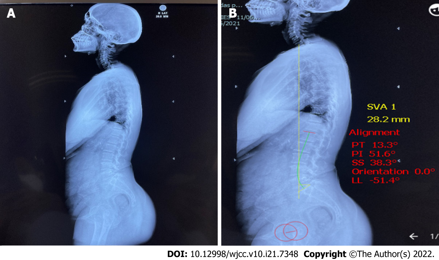
Figure 1 X-ray scannogram of the patient.
A: X-ray scannogram of whole spine without measurements; B: X-ray scannogram showing measurement in Surgimap software. LL: Lumbar lordosis; PI: Pelvic incidence; PT: Pelvic tilt; SS: Sacral slope; SVA: Sagittal vertical axis.
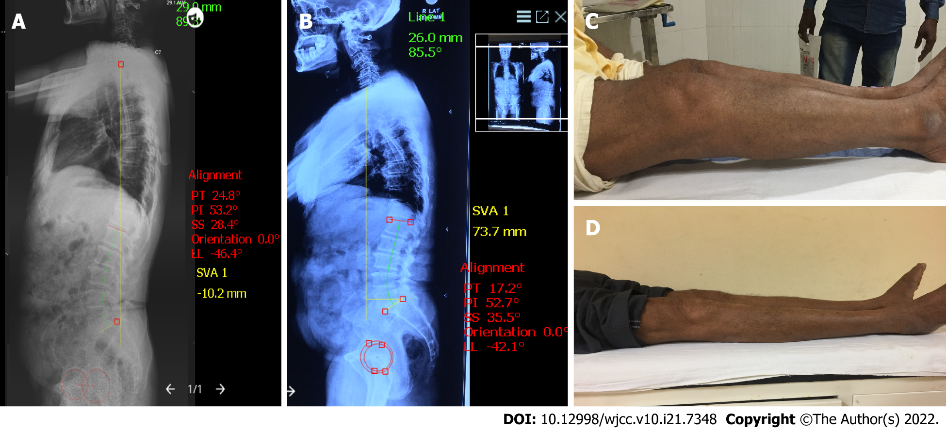
Figure 2 Complete clinical and radiological profile of the patient.
A: Preoperative scannogram showing the sagittal parameters; B: Postoperative scannogram showing the sagittal parameters; C: Preoperative clinical picture showing knee flexion deformity (KFD); D: Postoperative clinical picture showing correction of KFD.
- Citation: Puthiyapura LK, Jain M, Tripathy SK, Puliappadamb HM. Effect of osteoarthritic knee flexion deformity correction by total knee arthroplasty on sagittal spinopelvic alignment in Indian population. World J Clin Cases 2022; 10(21): 7348-7355
- URL: https://www.wjgnet.com/2307-8960/full/v10/i21/7348.htm
- DOI: https://dx.doi.org/10.12998/wjcc.v10.i21.7348


