Copyright
©The Author(s) 2022.
World J Clin Cases. Jul 26, 2022; 10(21): 7215-7223
Published online Jul 26, 2022. doi: 10.12998/wjcc.v10.i21.7215
Published online Jul 26, 2022. doi: 10.12998/wjcc.v10.i21.7215
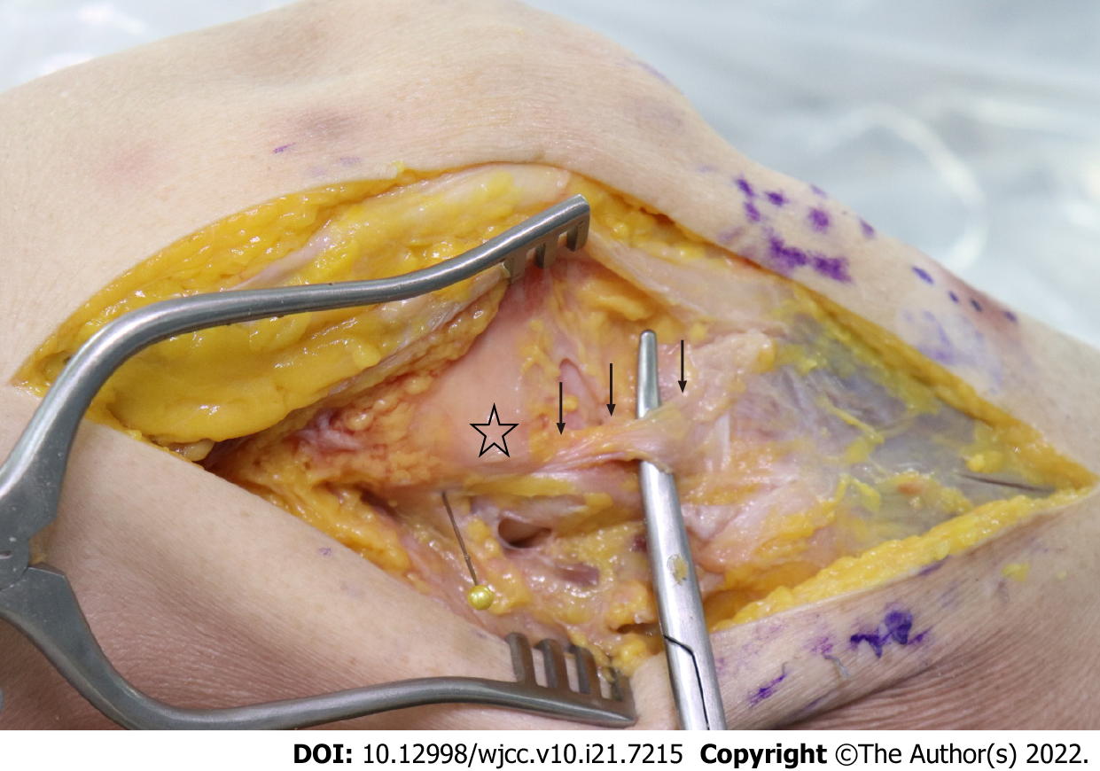
Figure 1 Photograph showing isolation of the anterolateral ligament (black arrows) in a cadaveric right knee joint.
The asterisk indicates the lateral epicondyle of the distal femur.
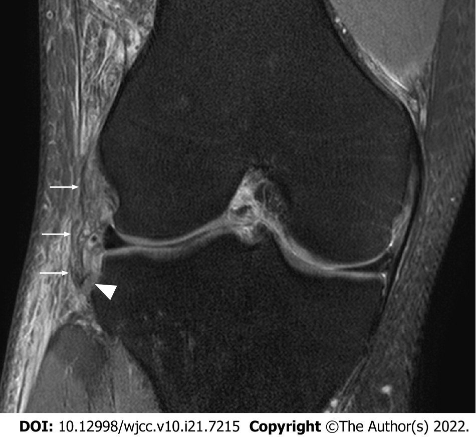
Figure 2 A coronal magnetic resonance image showing the anterolateral ligament (white arrows) which is attached to a Segond fracture fragment.
The white arrow head indicates a Segond fracture.
- Citation: Park JG, Han SB, Rhim HC, Jeon OH, Jang KM. Anatomy of the anterolateral ligament of the knee joint. World J Clin Cases 2022; 10(21): 7215-7223
- URL: https://www.wjgnet.com/2307-8960/full/v10/i21/7215.htm
- DOI: https://dx.doi.org/10.12998/wjcc.v10.i21.7215


