Copyright
©The Author(s) 2022.
World J Clin Cases. Jul 16, 2022; 10(20): 7147-7152
Published online Jul 16, 2022. doi: 10.12998/wjcc.v10.i20.7147
Published online Jul 16, 2022. doi: 10.12998/wjcc.v10.i20.7147
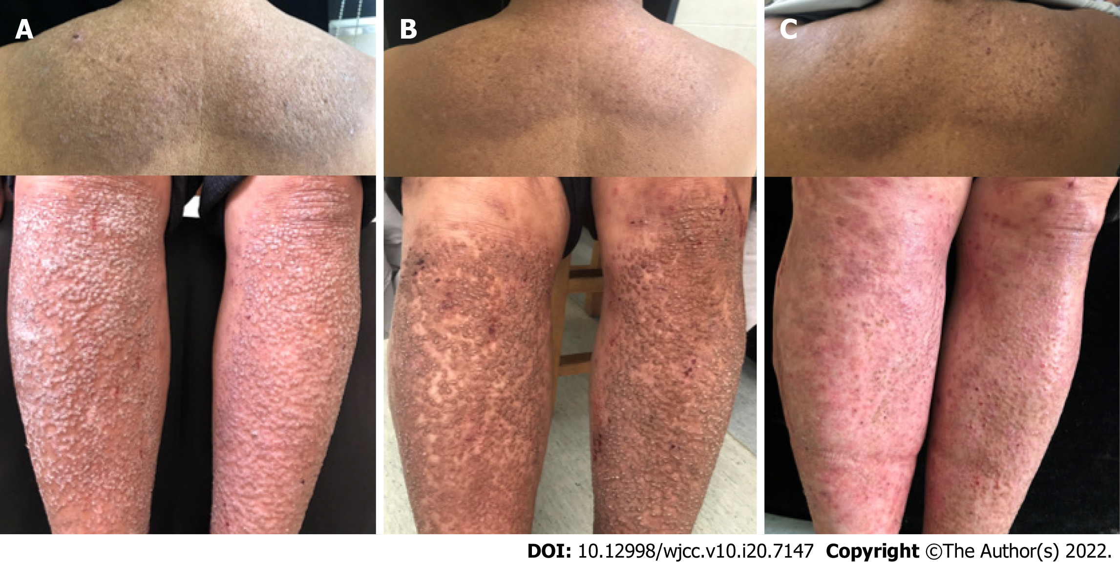
Figure 1 Close-up view of lesions in case 1.
A: Before treatment, the trunk and limbs had symmetrically distributed dense, brown, hard, and rough papules; B: After 4 wk of treatment, the lesions thinned; C: After 8 wk of treatment, the lesions disappeared and left residual pigmentation.
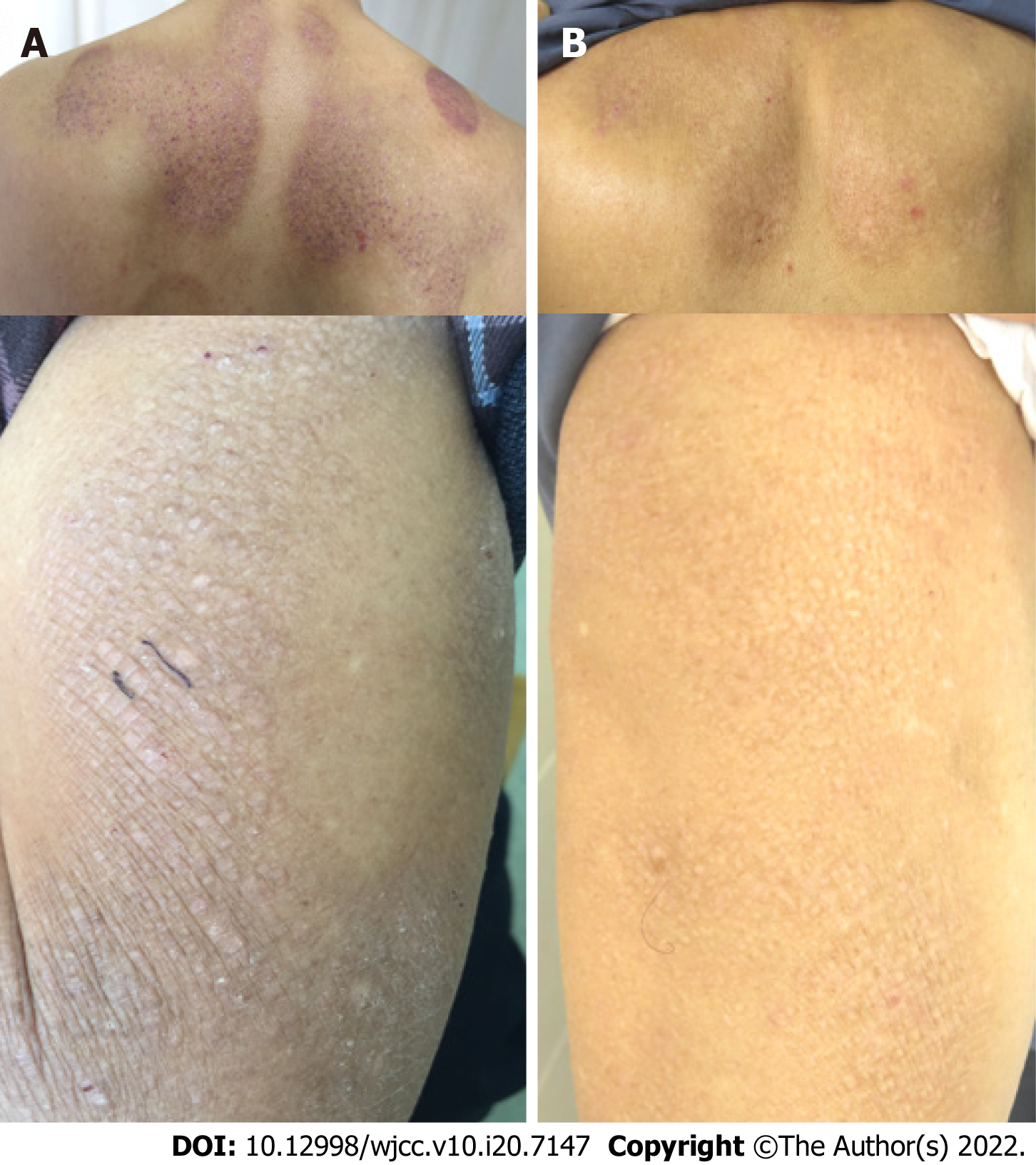
Figure 2 Close-up view of lesions in case 2.
A: Before treatment, polygonal mossy maculopapules were widely distributed in the dorsal scapular region and on both upper arms; B: After 4 wk of treatment, the lesions became flat with light brown pigmentation.
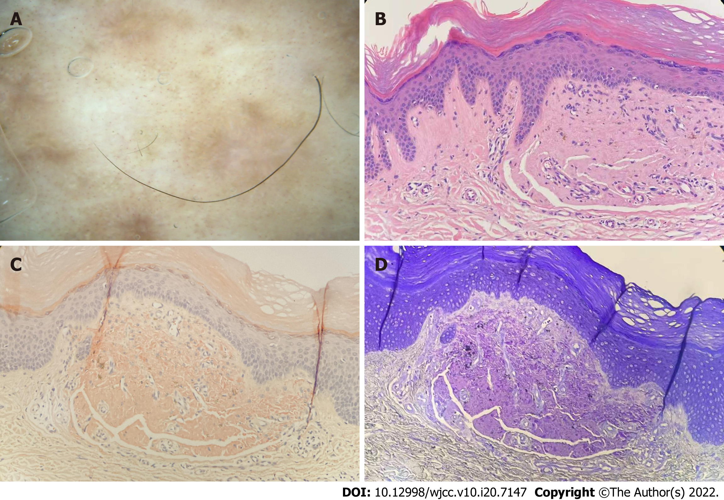
Figure 3 Dermatoscopic and dermatopathological findings in case 1.
A: The epidermis was hyperkeratinized. There were homogeneous red-stained amorphous substances in the dermis. Lymphocytes were scattered around the blood vessels; B: Amorphous substance was dyed orange; C: Amorphous substance was dyed purple; D: Dermatoscopy showed that the lesions were evenly distributed with gray pigmentation radially and dotted blood vessels densely distributed around them, and the central area was light white or light red without structure.
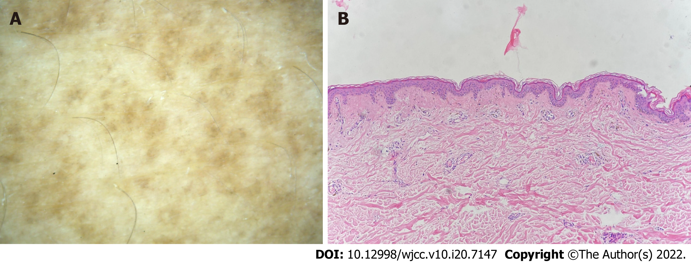
Figure 4 Dermatoscopic and dermatopathological findings of case 2.
A: In the dermal papillarylayer, there were a reddish mass of amorphous matter and pigment incontinence in the superficial dermis, and a small amount of lymphocyte infiltration around the blood vessels; B: Central hub had a white or brown structure, surrounded by irregular pigmentation.
- Citation: Su YQ, Liu ZY, Wei G, Zhang CM. Topical halometasone cream combined with fire needle pre-treatment for treatment of primary cutaneous amyloidosis: Two case reports. World J Clin Cases 2022; 10(20): 7147-7152
- URL: https://www.wjgnet.com/2307-8960/full/v10/i20/7147.htm
- DOI: https://dx.doi.org/10.12998/wjcc.v10.i20.7147


