Copyright
©The Author(s) 2022.
World J Clin Cases. Jul 16, 2022; 10(20): 7105-7115
Published online Jul 16, 2022. doi: 10.12998/wjcc.v10.i20.7105
Published online Jul 16, 2022. doi: 10.12998/wjcc.v10.i20.7105
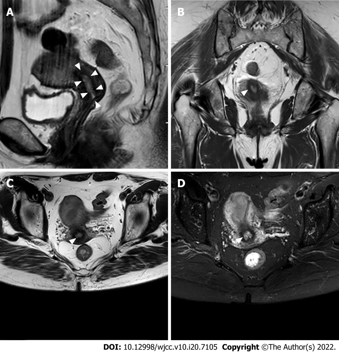
Figure 1 Preoperative abdominal magnetic resonance imaging.
A: Sagittal magnetic resonance imaging (MRI) showed an equal T1 and slightly longer T2 signal in the uterine cavity; B: Coronal MRI image; C: Axial MRI image. D: Enhanced MRI image showing that the lesions were inhomogeneous enhanced. The white arrowheads represent lesions. The tumor size was approximately 31 mm × 23 mm (B and C).
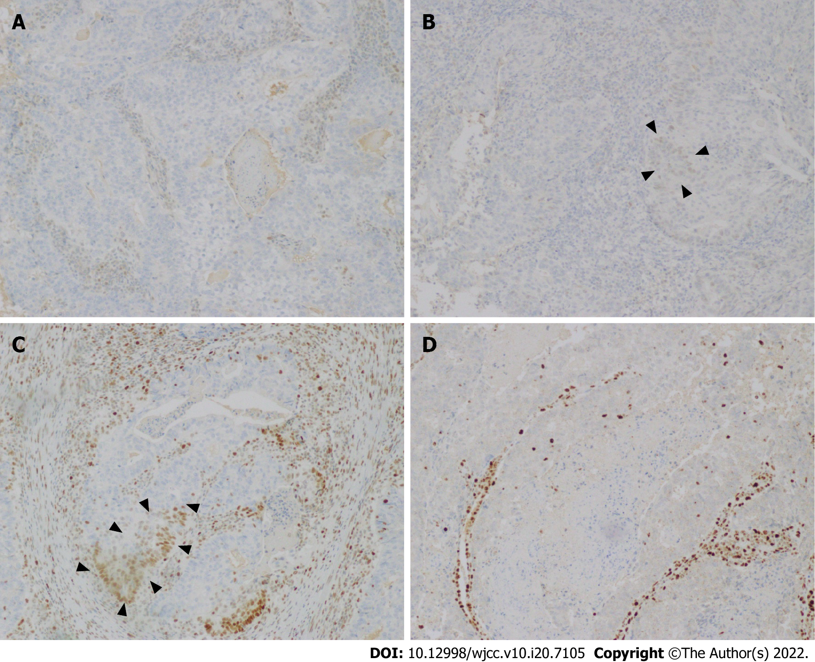
Figure 2 Immunohistochemical images.
A: Loss of MLH1 proteins was found in the tumor cells; B: Expression of MSH2 protein was detected in the tumor cells; C: Expression of MSH6 protein was detected in the tumor cells; D: Loss of PMS2 protein was found in the tumor cells.
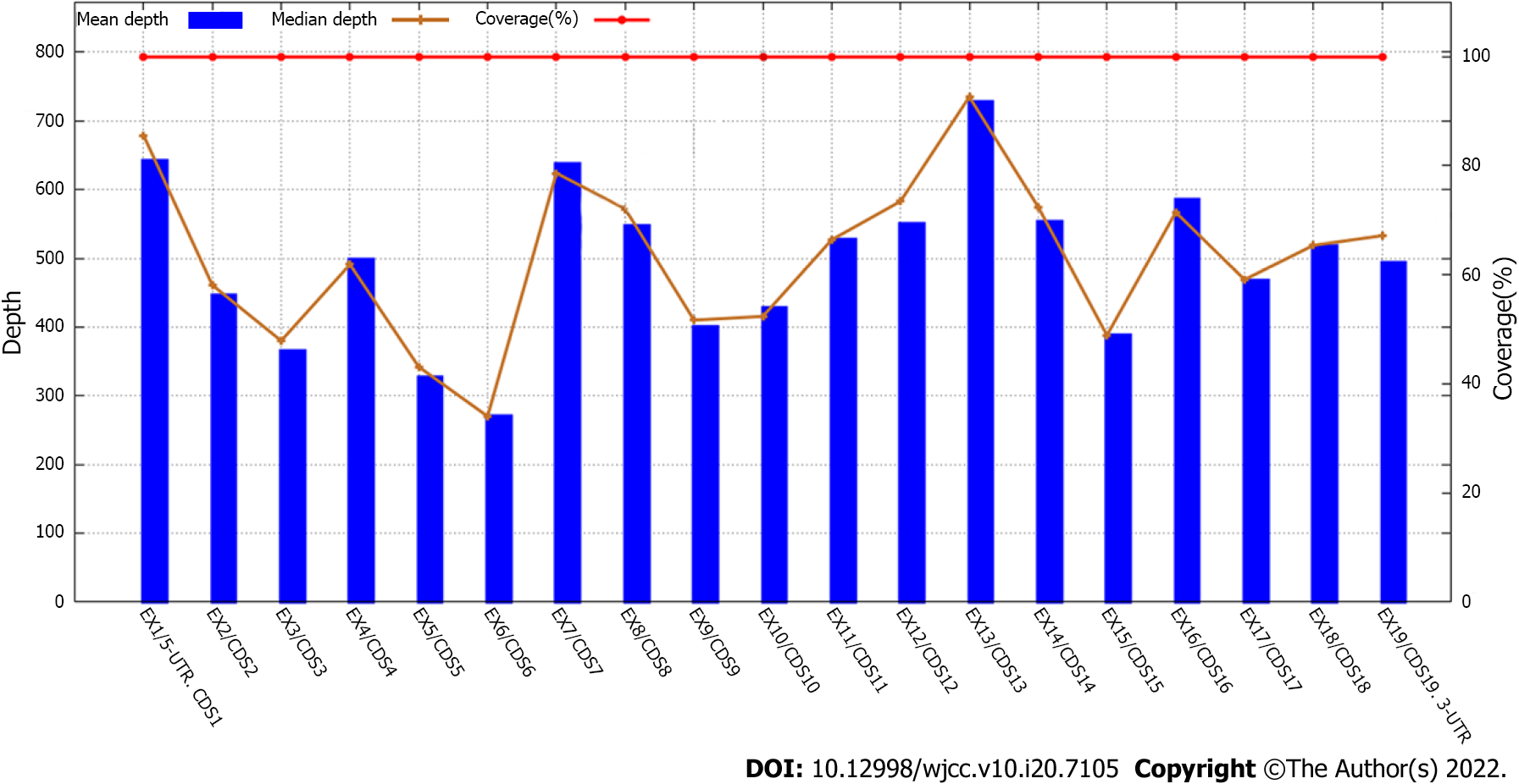
Figure 3 Figures related to gene test results.
A deletion mutation of exon 6 was found in the MLH1 gene.
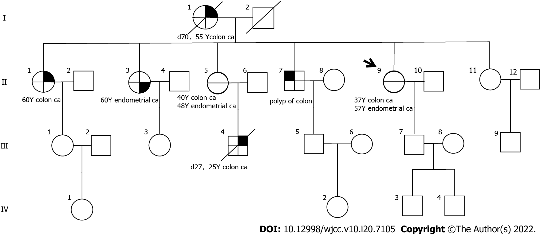
Figure 4 Family pedigree.
The reconstructed pedigree demonstrates that the proband (II-9), her mother (I-1), her sisters (II-1, II-3, and II-5) and her brother (II-7), and her sister’s son (IV-4) experienced cancer. I-1 was diagnosed with colon cancer at 55 years and died at 70 years. II-1 and II-3, both at 60 years, suffered from colon cancer and endometrial cancer, respectively. II-5 experienced colon cancer at 40 years and endometrial cancer at 48 years. II-7 developed polyps of the colon at unknown age. III-4 had colon cancer at 25 years and died at 27 years. The arrow indicates the proband. Solid symbols reveal persons affected by malignancy. The symbol with a slash indicates a deceased individual with age at death. Circles indicate female family members, and squares suggest male family members.
- Citation: Zhang XW, Jia ZH, Zhao LP, Wu YS, Cui MH, Jia Y, Xu TM. MutL homolog 1 germline mutation c.(453+1_454-1)_(545+1_546-1)del identified in lynch syndrome: A case report and review of literature. World J Clin Cases 2022; 10(20): 7105-7115
- URL: https://www.wjgnet.com/2307-8960/full/v10/i20/7105.htm
- DOI: https://dx.doi.org/10.12998/wjcc.v10.i20.7105


