Copyright
©The Author(s) 2022.
World J Clin Cases. Jul 16, 2022; 10(20): 7020-7028
Published online Jul 16, 2022. doi: 10.12998/wjcc.v10.i20.7020
Published online Jul 16, 2022. doi: 10.12998/wjcc.v10.i20.7020
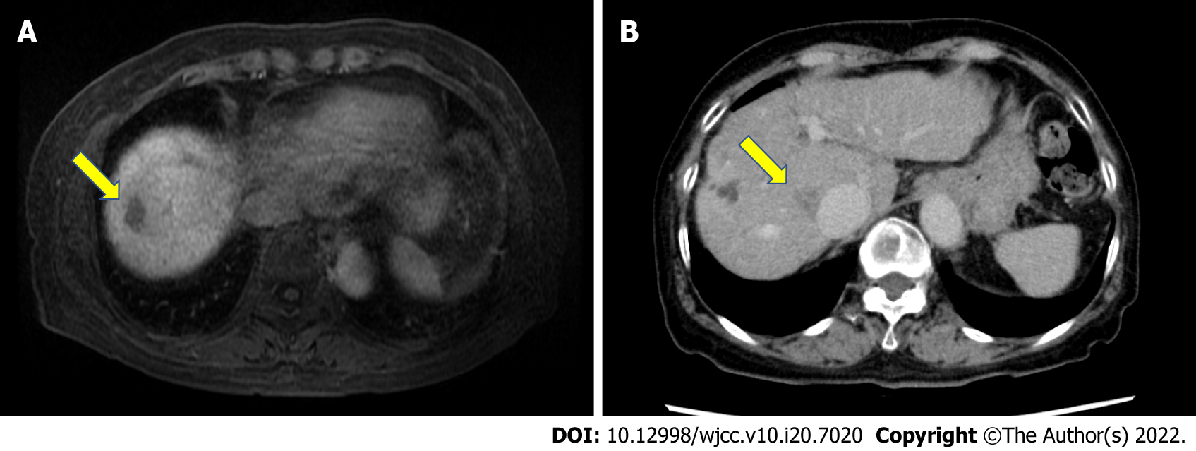
Figure 1 Location of tumors.
A: Gadolinium ethoxybenzyl diethylenetriamine pentaacetic acid-enhanced magnetic resonance imaging revealed a low-intensity area in segment VIII (S8) near the surface of the liver in the hepatobiliary phase (arrow); B: Abdominal contrast-enhanced computed tomography revealed a nodular lesion (20 mm) in S8 of the liver near the inferior vena cava, indicating washout in the delayed phase (arrow).
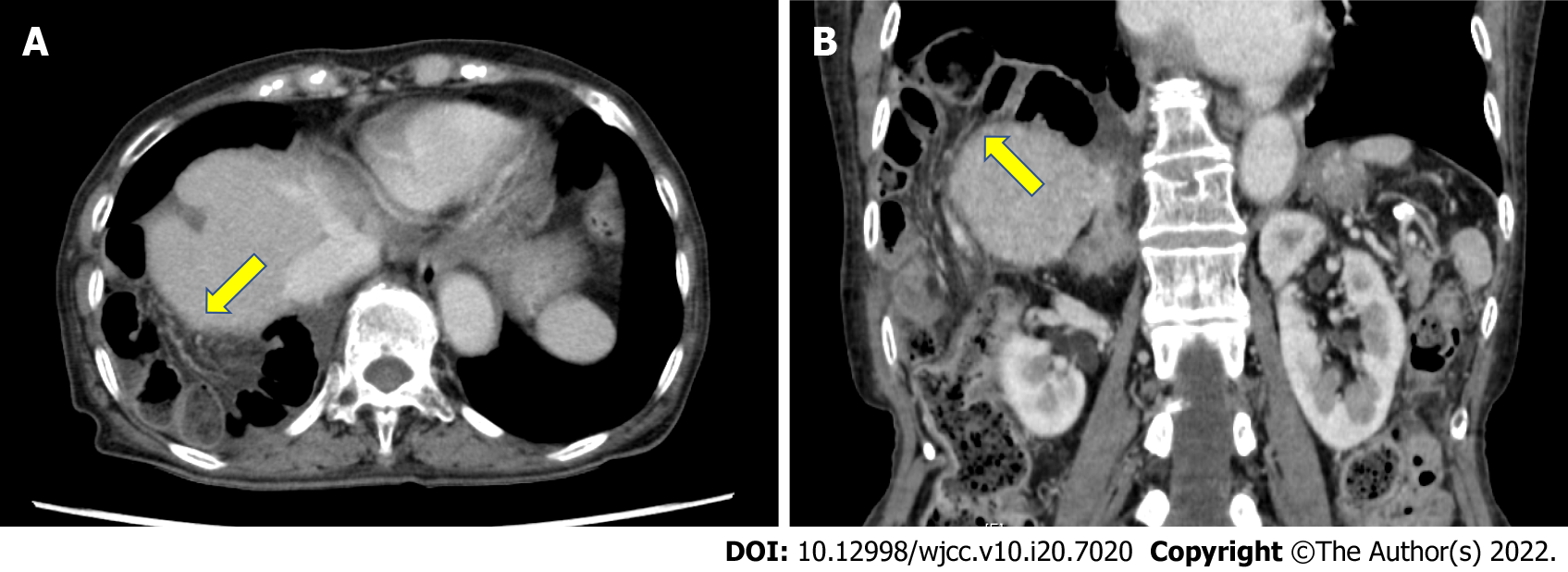
Figure 2 Contrast-enhanced computed tomography image at the onset of diaphragmatic hernia.
Contrast-enhanced CT revealed small intestine incarcerated in the right thorax (arrow). A: Horizontal plane; B: Coronal plane.
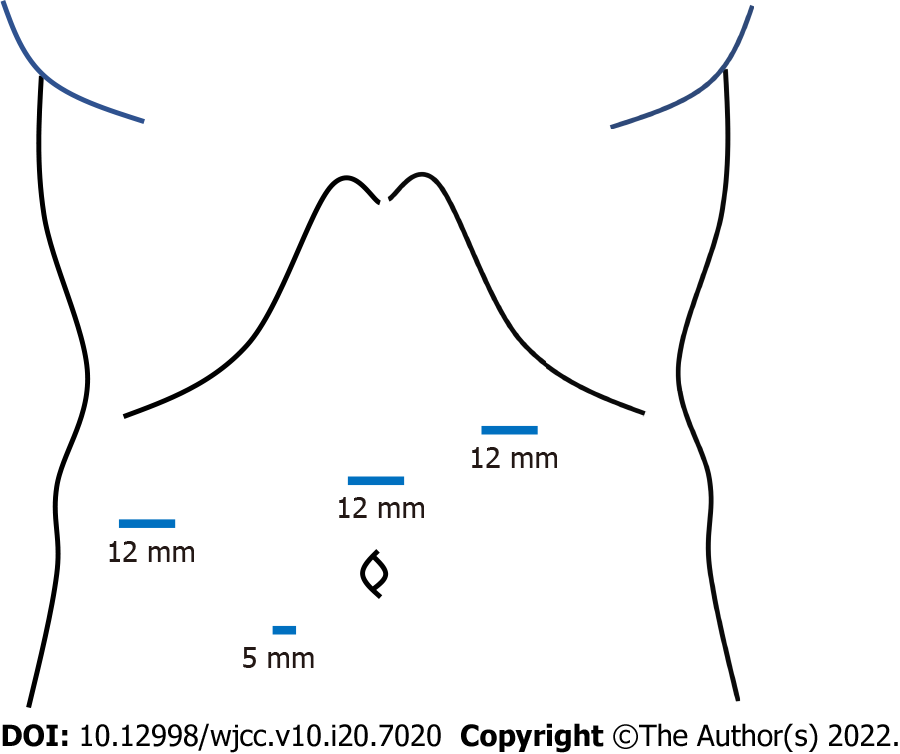
Figure 3 Scheme of trocars placement.
Four trocars were inserted into the abdomen. The first 12-mm trocar was introduced in the left-upper abdomen using the open-entry technique, while avoiding adhesions between the abdominal wall and visceral organs due to the previous surgery. After pneumoperitoneum by carbon dioxide insufflation, three more trocars were inserted at the right lateral abdomen, the mid-upper abdomen (12-mm trocars for operator) and near the umbilicus (a 5-mm trocar for scopist).
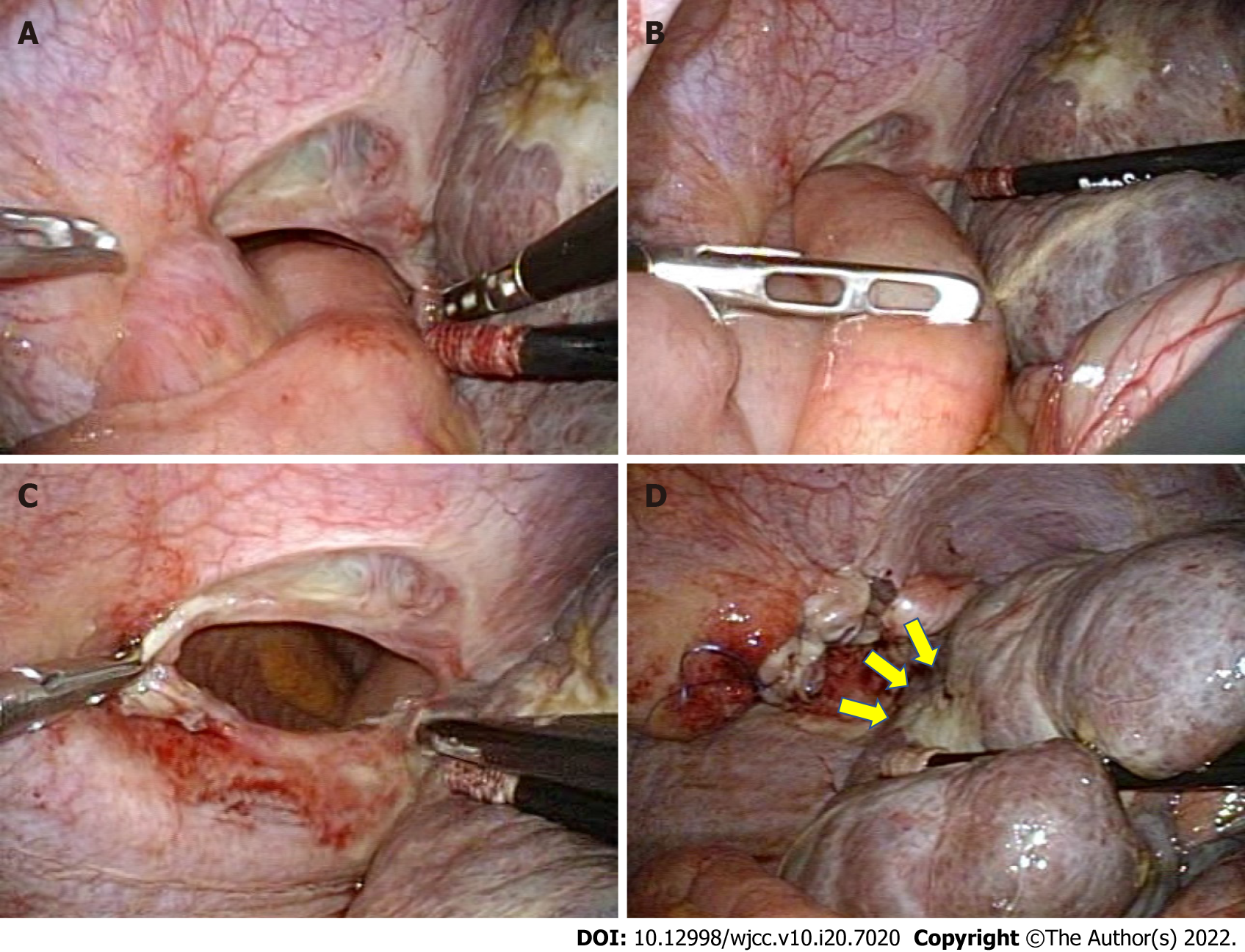
Figure 4 Surgical findings.
A: The small intestine had migrated through the diaphragmatic defect and was incarcerated in the right thorax; B: The small intestine was pulled back gently into the abdominal cavity by using laparoscopic bowel-grasping forceps; C: The size of the hernia orifice was estimated to be approximately 5 cm in diameter; D: The scar on the liver resulting from the first RFA was found to be close to the hernia opening (arrowheads). The defect was repaired using synthetic non-absorbable monofilament polypropylene sutures in the running fashion.
- Citation: Tsunoda J, Nishi T, Ito T, Inaguma G, Matsuzaki T, Seki H, Yasui N, Sakata M, Shimada A, Matsumoto H. Laparoscopic repair of diaphragmatic hernia associating with radiofrequency ablation for hepatocellular carcinoma: A case report. World J Clin Cases 2022; 10(20): 7020-7028
- URL: https://www.wjgnet.com/2307-8960/full/v10/i20/7020.htm
- DOI: https://dx.doi.org/10.12998/wjcc.v10.i20.7020


