Copyright
©The Author(s) 2022.
World J Clin Cases. Jul 16, 2022; 10(20): 6855-6864
Published online Jul 16, 2022. doi: 10.12998/wjcc.v10.i20.6855
Published online Jul 16, 2022. doi: 10.12998/wjcc.v10.i20.6855
Figure 1 Images of liver section plane.
A: Close-up image of the liver plane dissected using the water jet method. The fine Glissonian pedicles and veins were exposed without bleeding. There was no thermal damage to the resected surface; B: Image of the liver surface dissected with cavitron ultrasonic surgical aspirator (CUSA); C: Image of the liver surface dissected with the water jet. Thermal denaturation of the liver parenchyma with the water jet might be less than that with CUSA.
Figure 2 Measurement of computed tomography scan values.
A: Case 64 — water jet dissection: Computed tomography section of the portal venous phase with the maximum liver dissection length; B: Measurement of liver dissection length; C: Measurement of denaturation area of the cut surface.
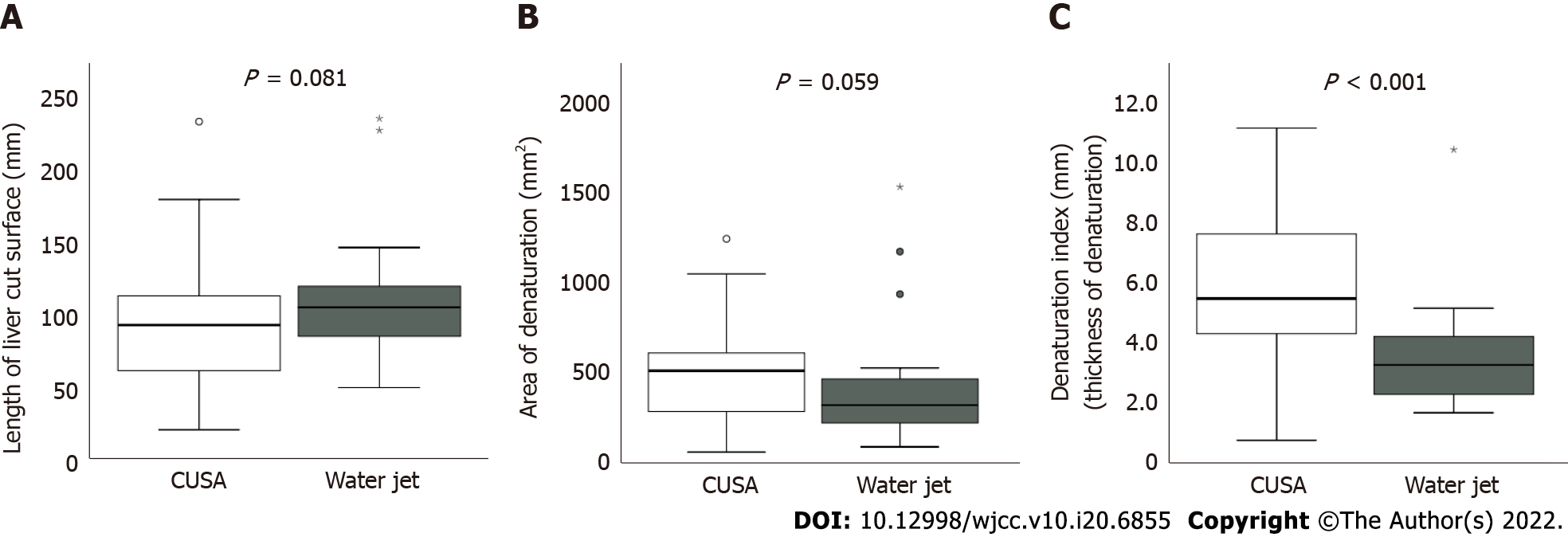
Figure 3 Comparison of computed tomography values between the two groups.
A: Length of the liver cut surface; B: Area of denaturation; C: Denaturation index (Mann-Whitney U test). CUSA: Cavitron ultrasonic surgical aspirator.
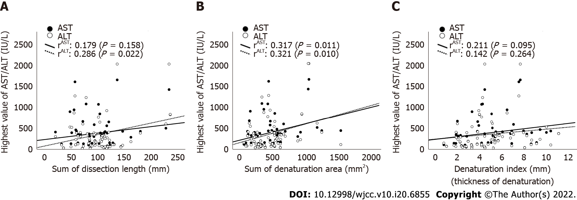
Figure 4 Correlation between the highest postoperative aspartate aminotransferase and alanine aminotransferase levels and computed tomography values.
A: Dissection length; B: Denaturation area; C: Denaturation index. There was a significant positive correlation between the highest aspartate aminotransferase/alanine aminotransferase levels and denaturation area. AST: Aspartate aminotransferase; ALT: Alanine aminotransferase.
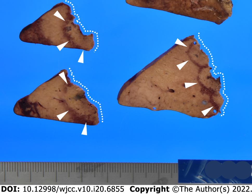
Figure 5 Example of the liver specimen (cut surface).
A thermal denaturation on a dissected plane of the liver; 5-8 mm of thermal denaturation (arrowhead) could be observed in the hepatic resection margin (dashed line) due to electrocauteries.
- Citation: Hanaki T, Tsuda A, Sunaguchi T, Goto K, Morimoto M, Murakami Y, Kihara K, Matsunaga T, Yamamoto M, Tokuyasu N, Sakamoto T, Hasegawa T, Fujiwara Y. Influence of the water jet system vs cavitron ultrasonic surgical aspirator for liver resection on the remnant liver. World J Clin Cases 2022; 10(20): 6855-6864
- URL: https://www.wjgnet.com/2307-8960/full/v10/i20/6855.htm
- DOI: https://dx.doi.org/10.12998/wjcc.v10.i20.6855


