Copyright
©The Author(s) 2022.
World J Clin Cases. Jan 14, 2022; 10(2): 717-724
Published online Jan 14, 2022. doi: 10.12998/wjcc.v10.i2.717
Published online Jan 14, 2022. doi: 10.12998/wjcc.v10.i2.717
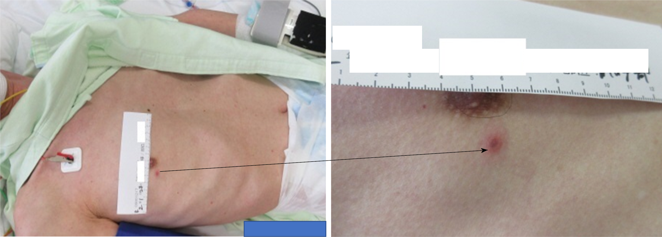
Figure 1 The skin lesion on the right chest.
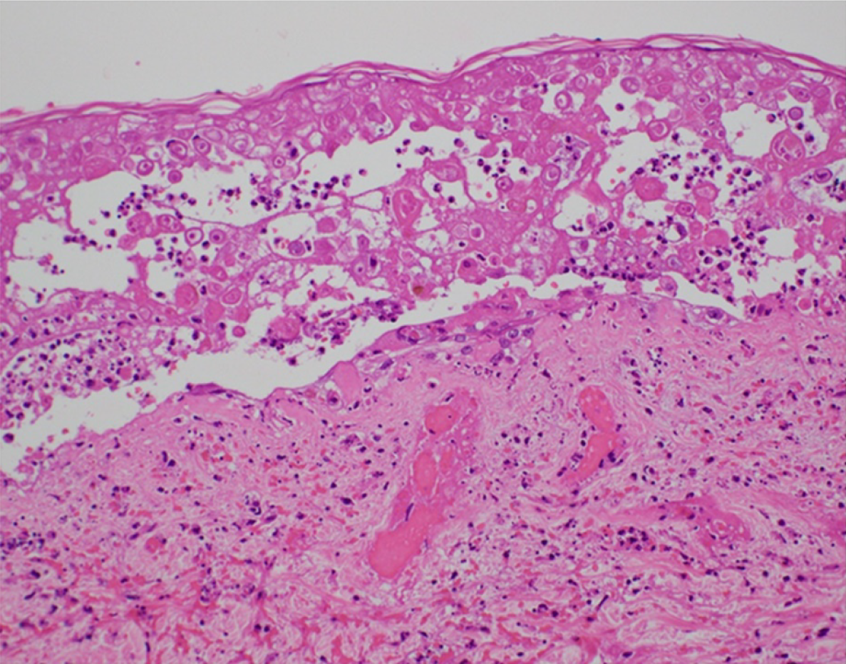
Figure 2 Skin biopsy of the lesion on the right chest.
The lesion shows intranuclear inclusion bodies and ballooning degeneration.
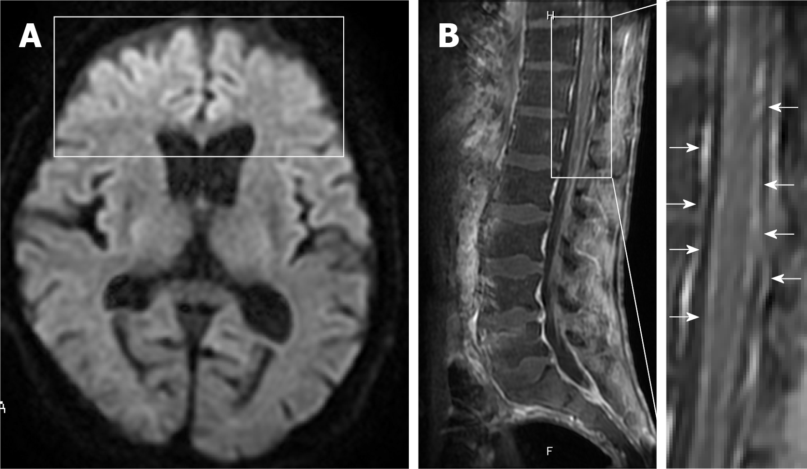
Figure 3 Axial diffusion-weighted magnetic resonance imaging on day 3 and sagittal contrast-enhanced T1-weighted magnetic resonance imaging on day 14.
A: The images show hyperintensities in the bilateral frontal lobes (block); B: Diffuse enhancement of meninges (arrows) with lumbar predominance and no cord enhancement.
- Citation: Takami K, Kenzaka T, Kumabe A, Fukuzawa M, Eto Y, Nakata S, Shinohara K, Endo K. Varicella-zoster virus-associated meningitis, encephalitis, and myelitis with sporadic skin blisters: A case report. World J Clin Cases 2022; 10(2): 717-724
- URL: https://www.wjgnet.com/2307-8960/full/v10/i2/717.htm
- DOI: https://dx.doi.org/10.12998/wjcc.v10.i2.717


