Copyright
©The Author(s) 2022.
World J Clin Cases. Jan 14, 2022; 10(2): 691-702
Published online Jan 14, 2022. doi: 10.12998/wjcc.v10.i2.691
Published online Jan 14, 2022. doi: 10.12998/wjcc.v10.i2.691
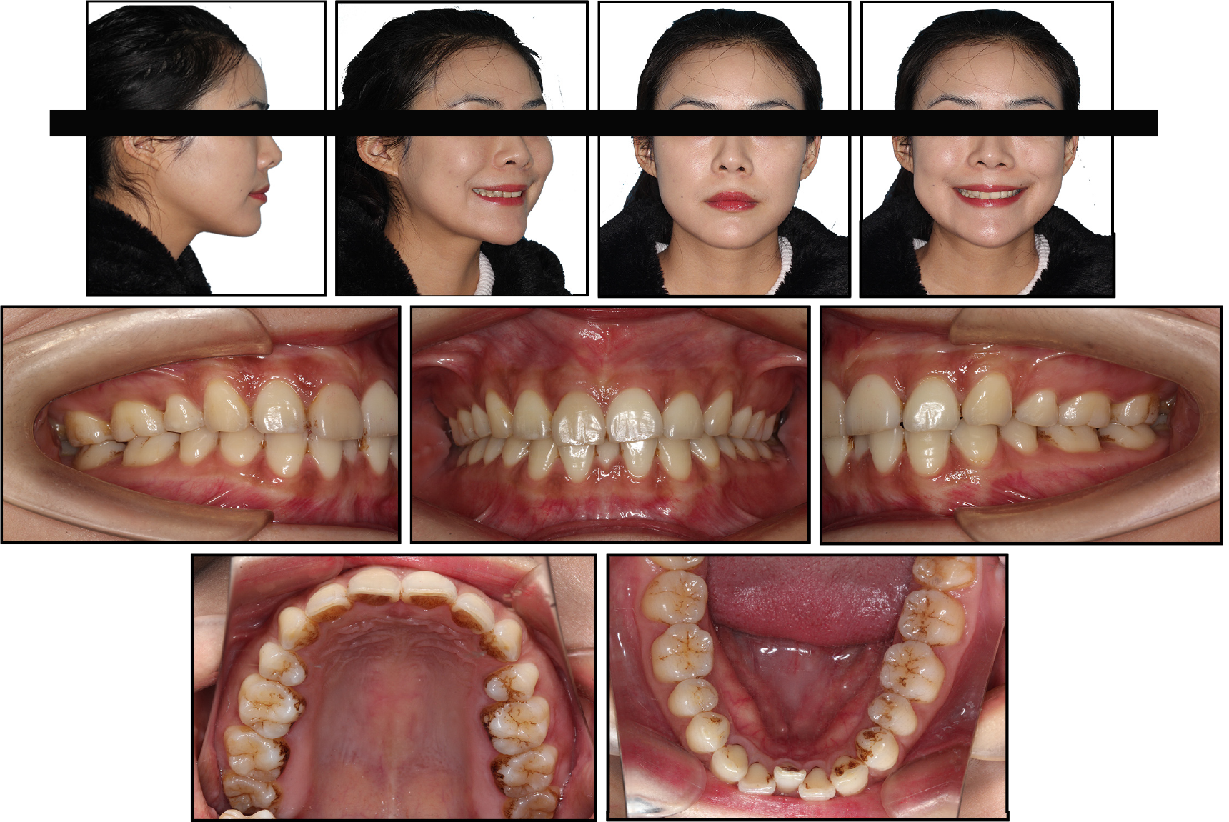
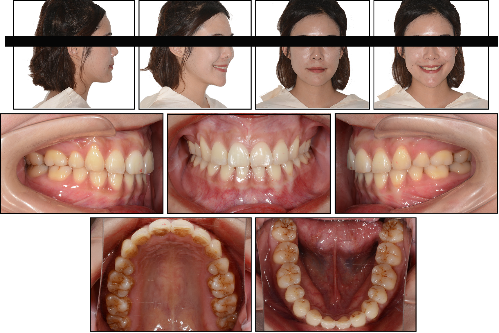
Figure 1 Pretreatment intraoral and facial photographs.
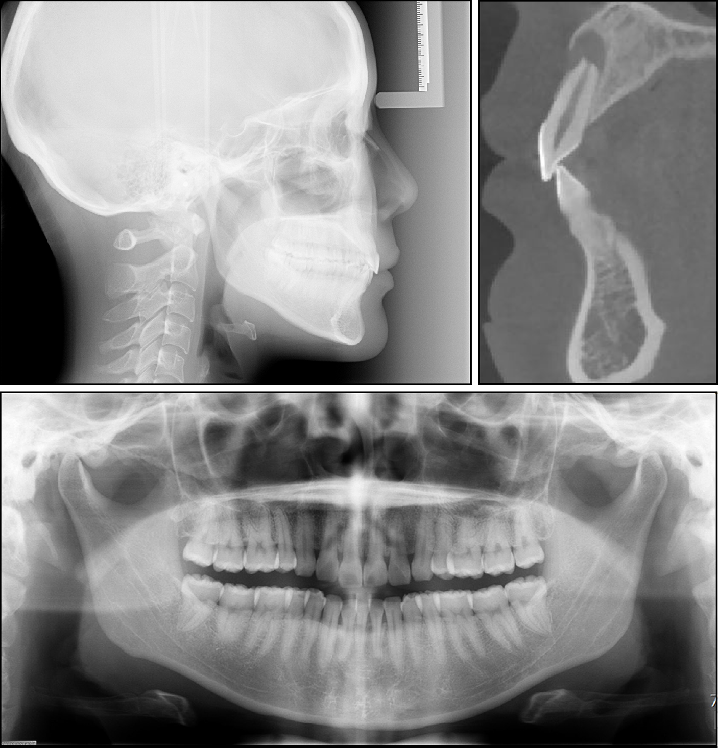
Figure 2 Pretreatment radiographs and cone beam computed tomography image of the upper right central incisor.
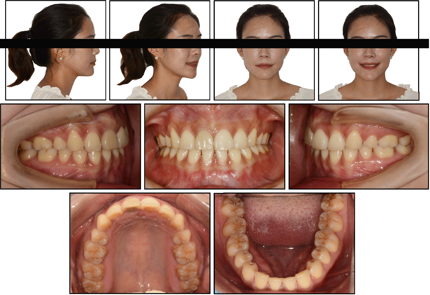
Figure 3 Posttreatment intraoral and facial photographs.
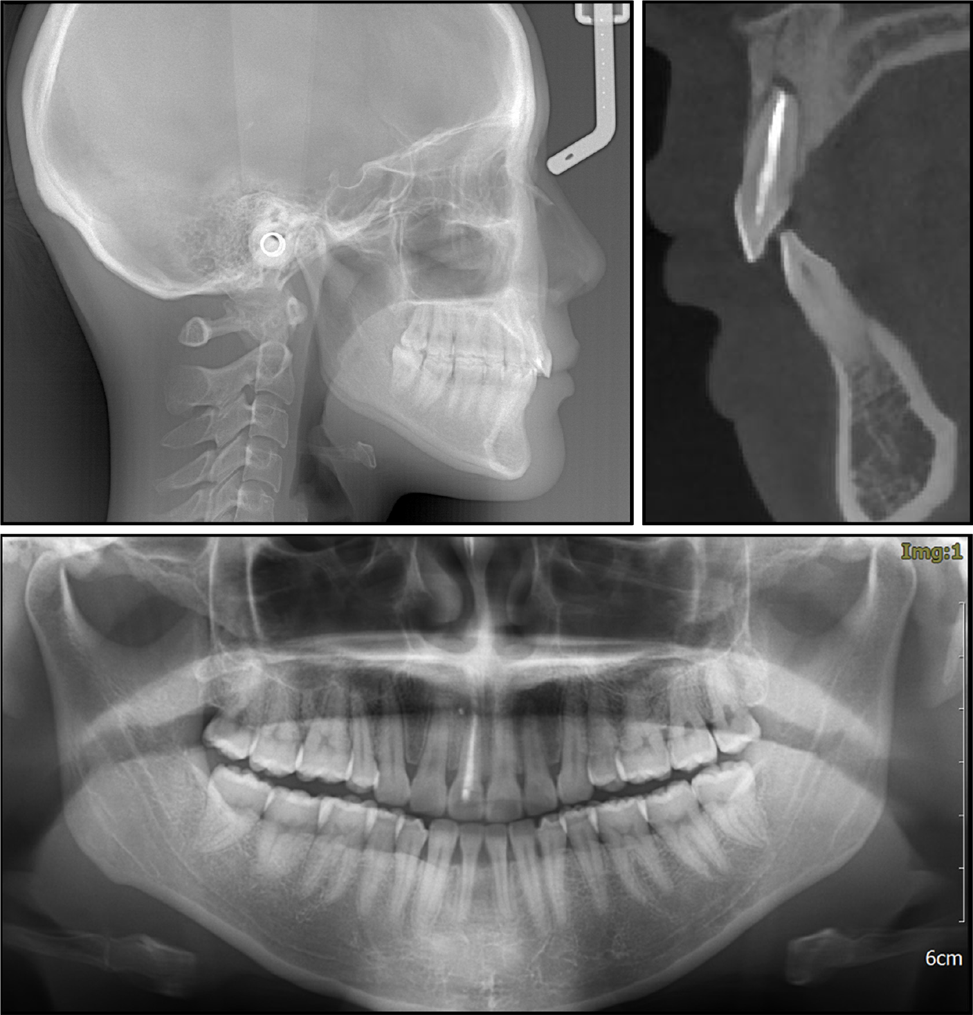
Figure 4 Posttreatment radiographs and cone beam computed tomography image of the upper right central incisor.
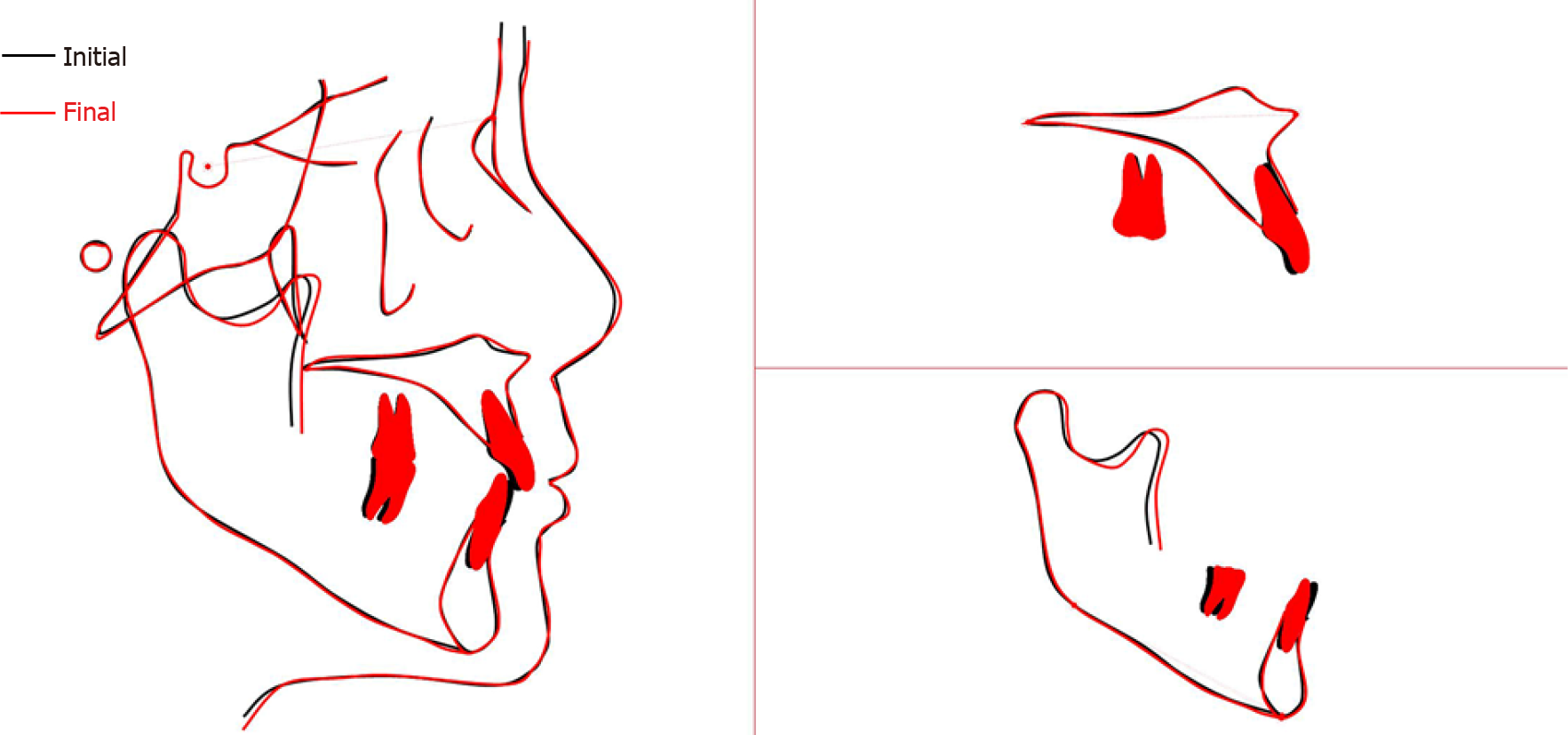
Figure 5 Superimposition of pretreatment (black) and posttreatment (red) cephalometric tracings.
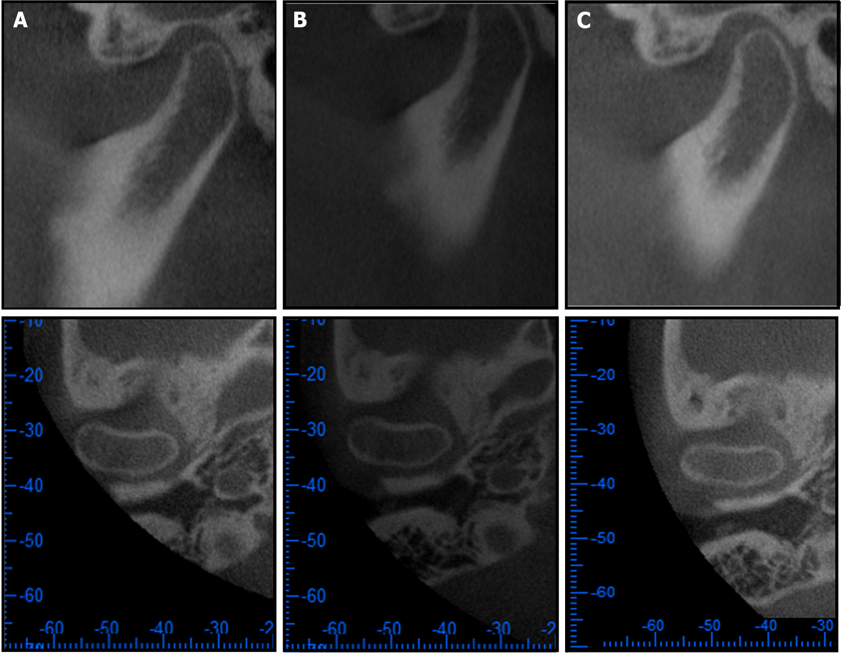
Figure 6 Cone beam computed tomography images of the right TMJ in the sagittal (upper) and transverse (lower) planes.
A: Pretreatment; B: Posttreatment; C: 22-mo retention.
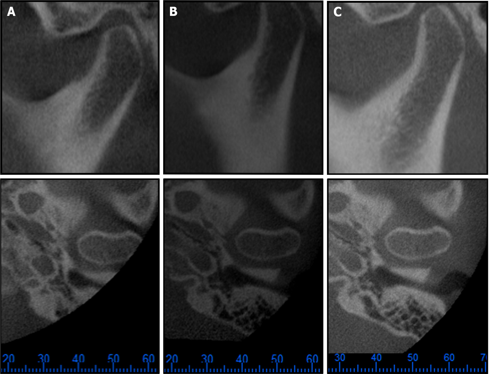
Figure 7 Cone beam computed tomography images of the left TMJ in the sagittal (upper) and transverse (lower) planes.
A: Pretreatment; B: Posttreatment; C: 22-mo retention.
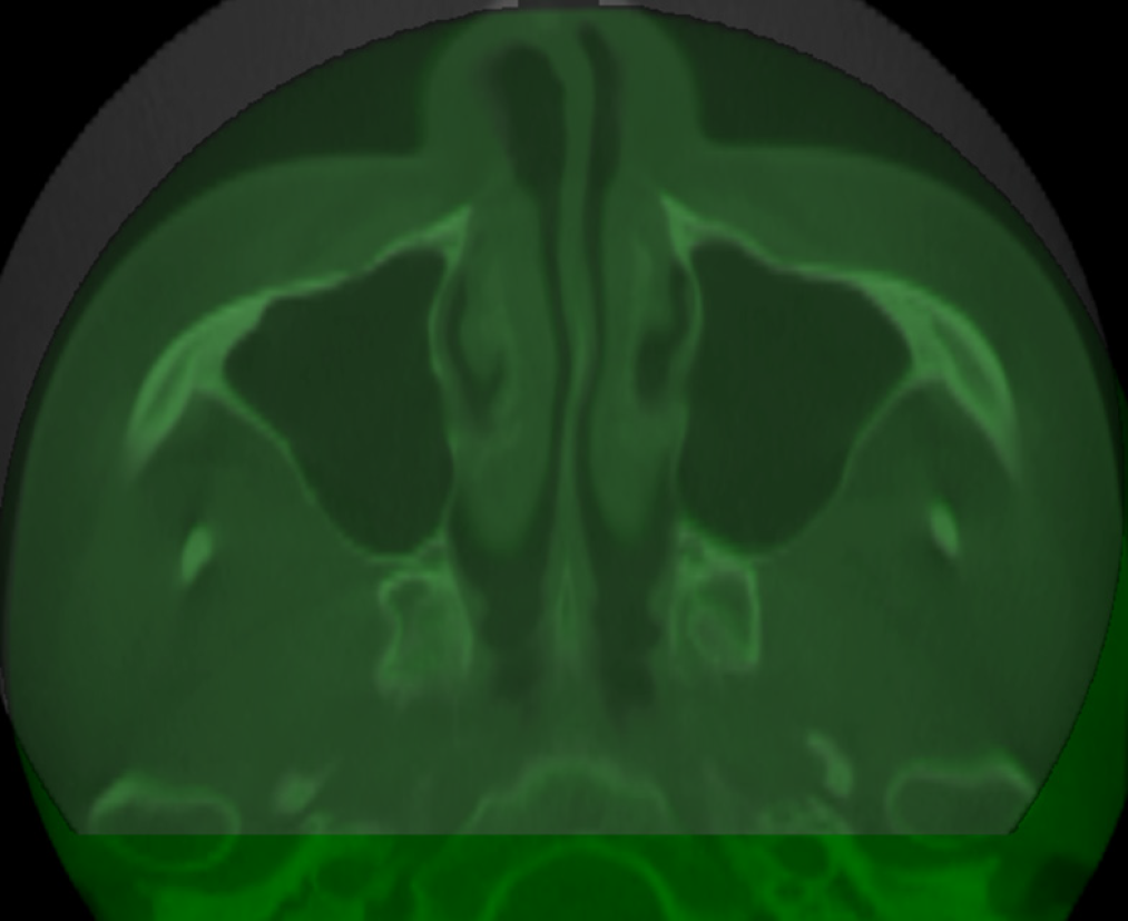
Figure 8 Cone beam computed tomography superimposition of pretreatment (gray) and 22-mo retention (green) bilateral temporomandibular joints.
Figure 9 Intraoral and facial photographs after 22 mo of retention.
- Citation: Yu LY, Xia K, Sun WT, Huang XQ, Chi JY, Wang LJ, Zhao ZH, Liu J. Orthodontic retreatment of an adult woman with mandibular backward positioning and temporomandibular joint disorder: A case report. World J Clin Cases 2022; 10(2): 691-702
- URL: https://www.wjgnet.com/2307-8960/full/v10/i2/691.htm
- DOI: https://dx.doi.org/10.12998/wjcc.v10.i2.691


