Copyright
©The Author(s) 2022.
World J Clin Cases. Jan 14, 2022; 10(2): 663-670
Published online Jan 14, 2022. doi: 10.12998/wjcc.v10.i2.663
Published online Jan 14, 2022. doi: 10.12998/wjcc.v10.i2.663
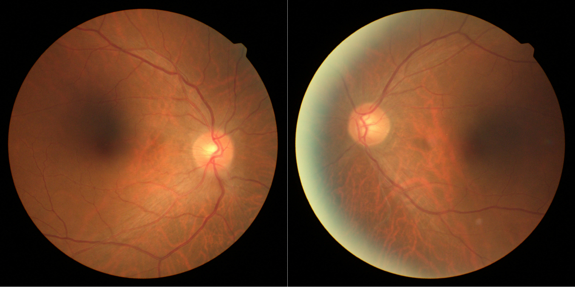
Figure 1 Color fundus photography at the initial visit.
The optic disk of both eyes was normal at the initial visit.
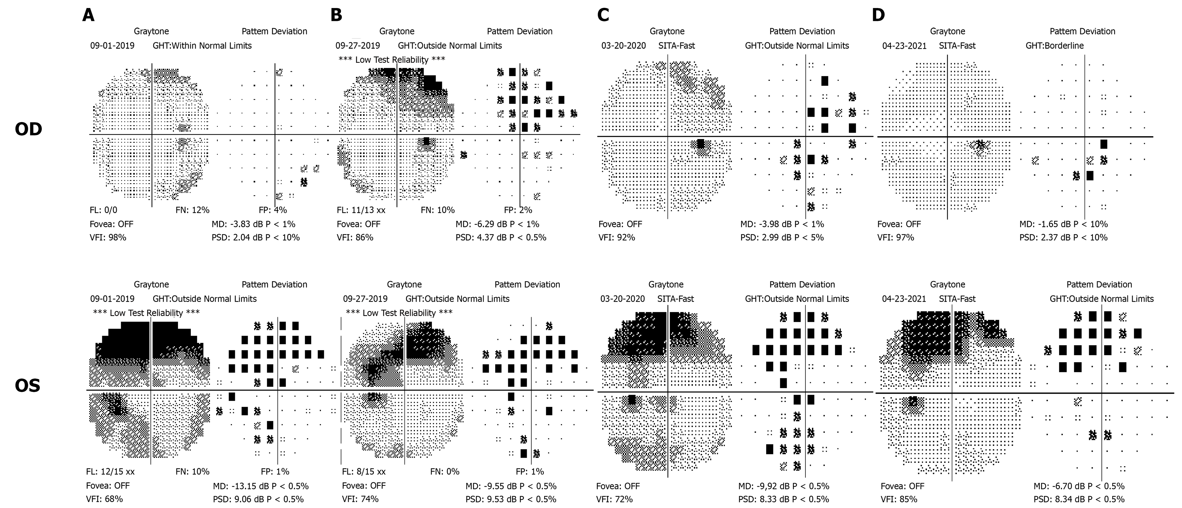
Figure 2 Computer perimetry results of four follow-up visits.
A: At the initial visit, there were suspicious dark spots in the inferonasal region of the right eye, and the visual field defect of the left eye manifested as concentric contraction; B: At the second visit, visual field defects had worsened in the right eye; C: At the third visit, the defect showed progression in the left eye, but improved in the right eye; D: At the fourth visit, the visual field in both eyes improved.
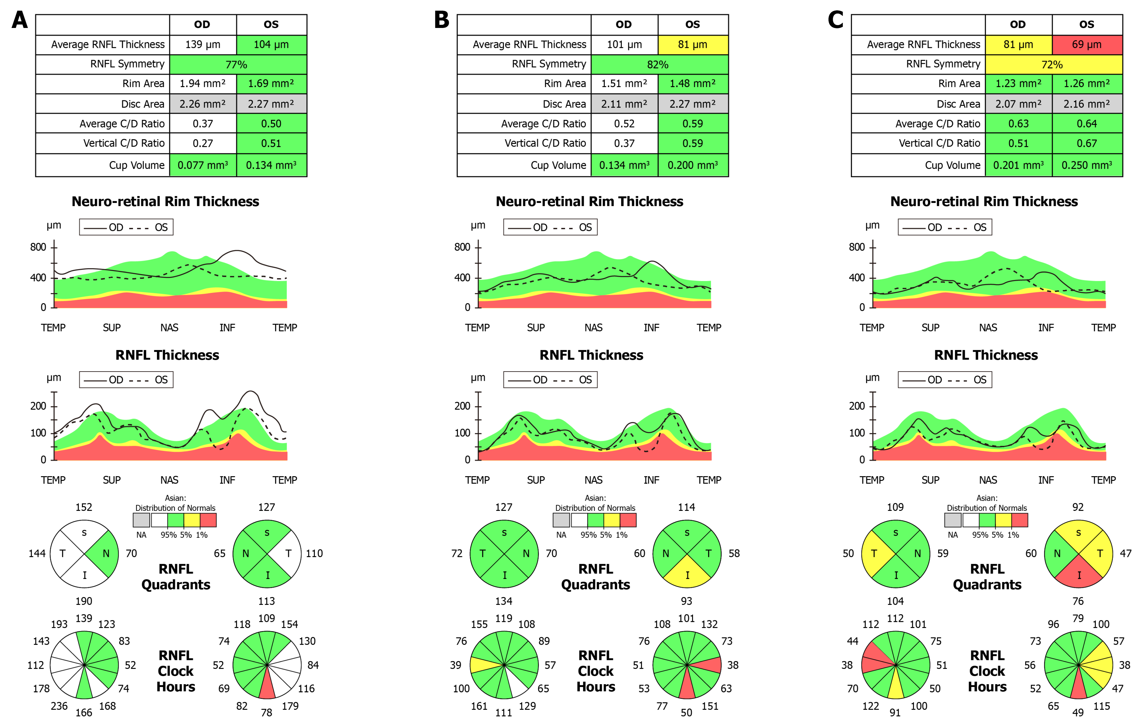
Figure 3 Retinal nerve fiber layer high-definition optical coherence tomography.
A: At the first visit, the retinal nerve fiber layer (RNFL) of the right eye had mild thickening in the superior, inferior and nasal regions; the RNFL of the left eye had mild thickening on the temporal side; B: Six months after drug withdrawal, the RNFL of both eyes became thinner compared with the first visit; the RNFL of the right eye was within the normal range; and the average and inferior thicknesses of the RNFL in the left eye were lower than normal; C: Eighteen months after stopping drug treatment, the RNFL of both eyes further decreased, and the temporal side of both eyes became thinner significantly.
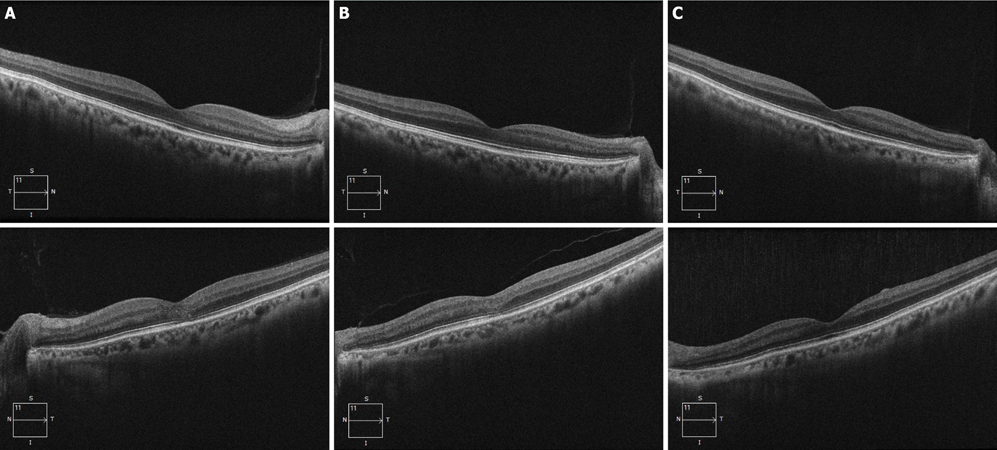
Figure 4 Outer nuclear layer high-definition optical coherence tomography.
A: At the initial visit, the outer nuclear layer under the fovea and the outer reflection bands representing the photoreceptor cells of the left eye were blurred (mainly the ellipsoid zone and the intersection area), and the damaged intersection area was mainly on the nasal side of the macula; B: Six months after drug withdrawal, compared with the first visit, the structure of the outer nuclear layer and ellipsoid zone of the macula had partially recovered; C: Eighteen months after drug withdrawl, the structure of the outer nuclear layer and the ellipsoid zone of the macula had recovered to normal appearance.
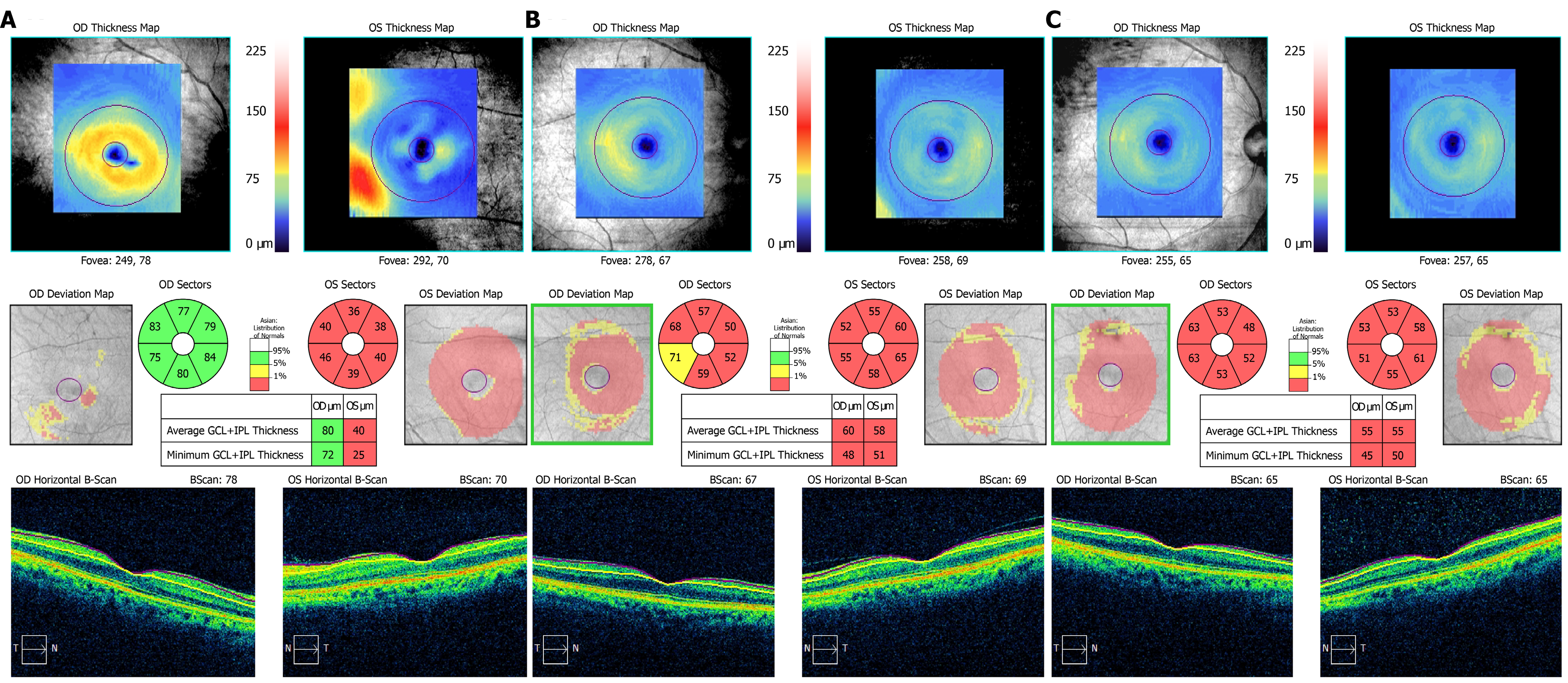
Figure 5 Ganglion cell layer and inner plexiform layer high-definition optical coherence tomography.
A: At the first visit, the thickness of ganglion cell layer and inner plexiform layer (GCIPL) in the right eye was normal, and the GCIPL in the left eye was significantly lower than normal; B: At the second visit, the GCIPL of the right eye also decreased; C: At the third visit, the binocular visual function recovered, but the GCIPL was still lower than normal without any obvious recovery.
- Citation: Sheng WY, Wu SQ, Su LY, Zhu LW. Ethambutol-induced optic neuropathy with rare bilateral asymmetry onset: A case report. World J Clin Cases 2022; 10(2): 663-670
- URL: https://www.wjgnet.com/2307-8960/full/v10/i2/663.htm
- DOI: https://dx.doi.org/10.12998/wjcc.v10.i2.663


