Copyright
©The Author(s) 2022.
World J Clin Cases. Jan 14, 2022; 10(2): 656-662
Published online Jan 14, 2022. doi: 10.12998/wjcc.v10.i2.656
Published online Jan 14, 2022. doi: 10.12998/wjcc.v10.i2.656
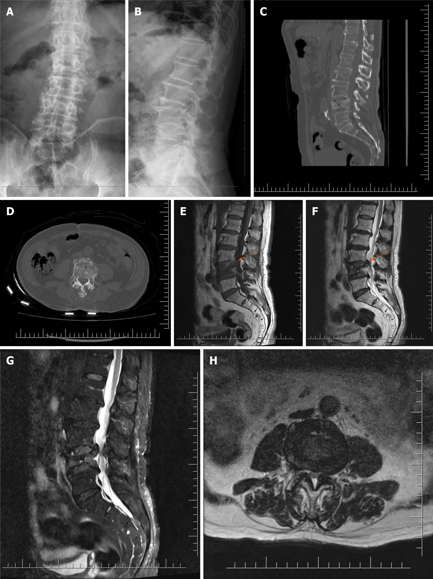
Figure 1 Imaging examination before percutaneous vertebroplasty.
A: Anteroposterior X-ray before percutaneous vertebroplasty (PVP); B: Lateral X-ray before PVP; C: Sagittal computed tomography (CT) reconstruction before PVP; D: CT cross- section reconstruction before PVP; E: T1 magnetic resonance imaging (MRI) before PVP; F: T2 MRI before PVP; G: T2 fat suppression MRI before PVP; H: T2 cross section MRI before PVP.
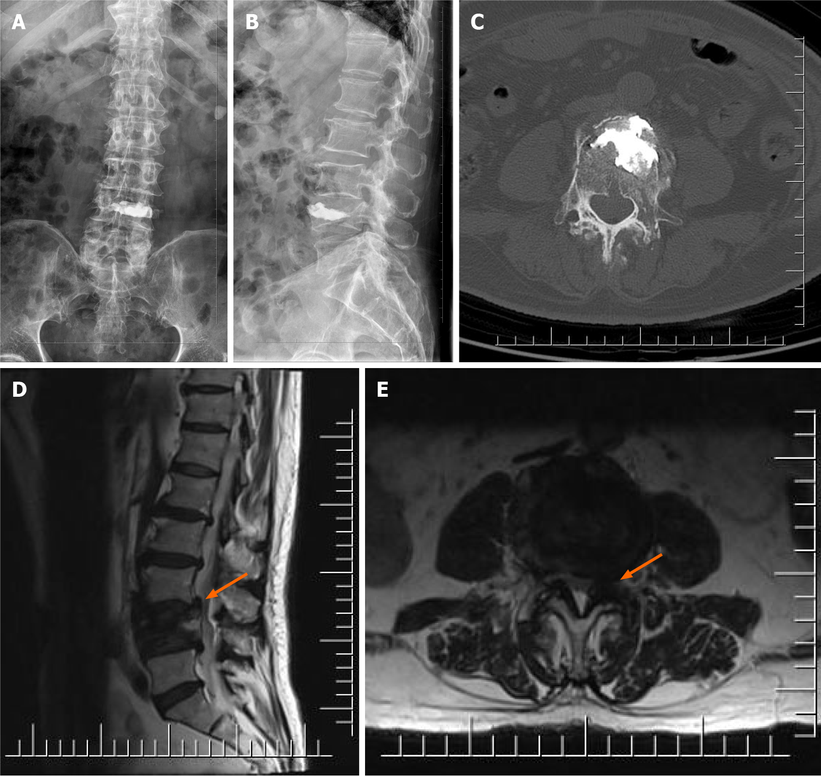
Figure 2 Imaging examination after percutaneous vertebroplasty.
A: Anteroposterior X-ray after percutaneous vertebroplasty (PVP); B: Lateral X-ray after PVP; C: Computed tomography cross section after PVP; D: T2 of magnetic resonance imaging (MRI) after PVP; E: T2 cross section of MRI after PVP.
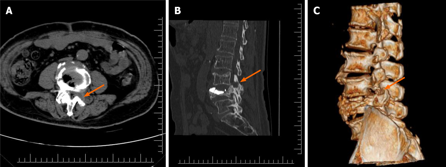
Figure 3 Imaging examination after full-endoscopic spine surgery.
A: Computed tomography (CT) cross-section after full-endoscopic spine surgery (FESS); B: Sagittal CT reconstruction after FESS; C: Three-dimensional CT reconstruction after FESS.
- Citation: Zhao QL, Hou KP, Wu ZX, Xiao L, Xu HG. Full-endoscopic spine surgery treatment of lumbar foraminal stenosis after osteoporotic vertebral compression fractures: A case report. World J Clin Cases 2022; 10(2): 656-662
- URL: https://www.wjgnet.com/2307-8960/full/v10/i2/656.htm
- DOI: https://dx.doi.org/10.12998/wjcc.v10.i2.656


