Copyright
©The Author(s) 2022.
World J Clin Cases. Jan 14, 2022; 10(2): 643-655
Published online Jan 14, 2022. doi: 10.12998/wjcc.v10.i2.643
Published online Jan 14, 2022. doi: 10.12998/wjcc.v10.i2.643
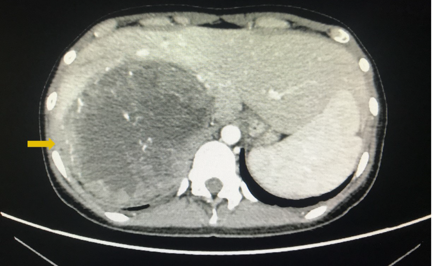
Figure 1 Retroperitoneal mass detected on enhanced abdominal computed tomography.
The yellow arrow indicates the tumor was closely related to the liver.
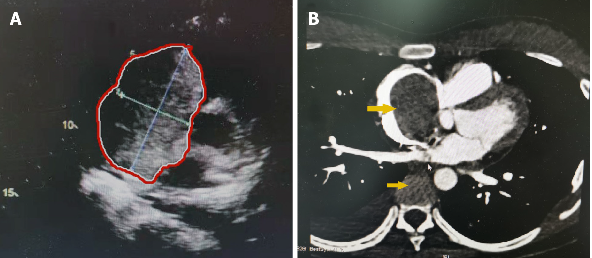
Figure 2 Tumor thrombus detected in the inferior vena cava and right atrium.
A: Preoperative transthoracic echocardiography showed that the tumor thrombus extended into the right atrium. Red areas indicate tumor thrombus; B: Cardiac enhanced magnetic resonance imaging showed tumor thrombus in the inferior vena cava (IVC) and the right atrium. The upper yellow arrow indicates tumor thrombus in the right atrium. The lower yellow arrow indicates tumor thrombus in the IVC.
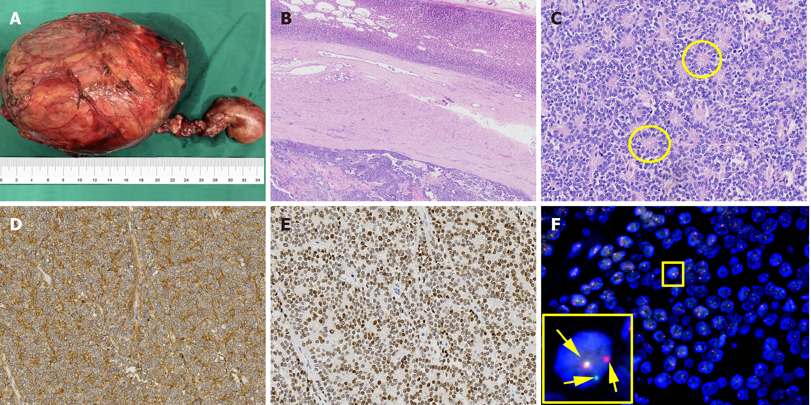
Figure 3 Pathological findings of the resected primary tumor.
A: Image of the resected tumor and thrombus; B: Hematoxylin-Eosin staining shows that the tumor (the lower part of the image) was surrounded by normal adrenal tissue (the upper part of the image), which suggested that the tumor arose in the adrenal gland (2 × magnification); C: Hematoxylin-Eosin staining shows the tumor cells with round nuclei and pale cytoplasm, as well as their rosette structures (inside the yellow circles) (20 × magnification); D: Immunohistochemistry showed CD99 positive tumor cells (20 × magnification); E: Immunohistochemistry showed Nkx2.2 positive tumor cells (20 × magnification); F: Separation of the red signal and green signal (the right two signals) by fluorescence in situ hybridization reveals Ewing’s sarcoma breakpoint region 1 gene rearrangement in the nuclei of tumor cells, while the yellow signal (the left signal) shows the normal allele.
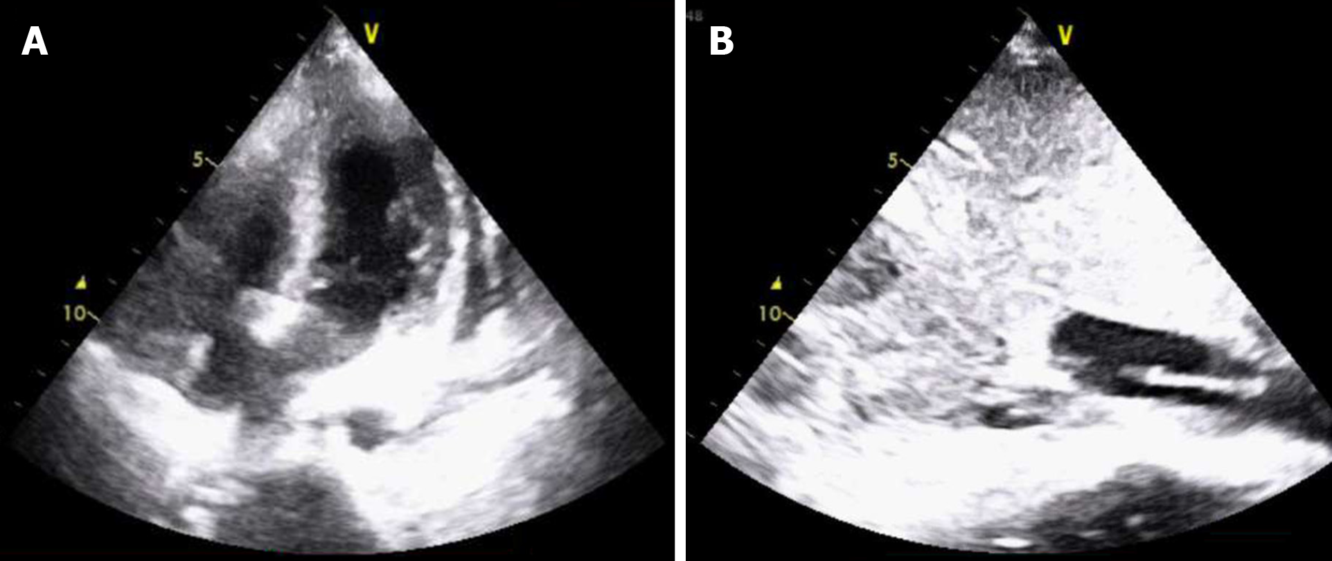
Figure 4 Echocardiogram.
A: The postoperative transthoracic echocardiography examination in the intensive care unit showed that there was no obvious tumor thrombus in the right atrium; B: There was a 7 cm diameter mass in the inferior vena cava.
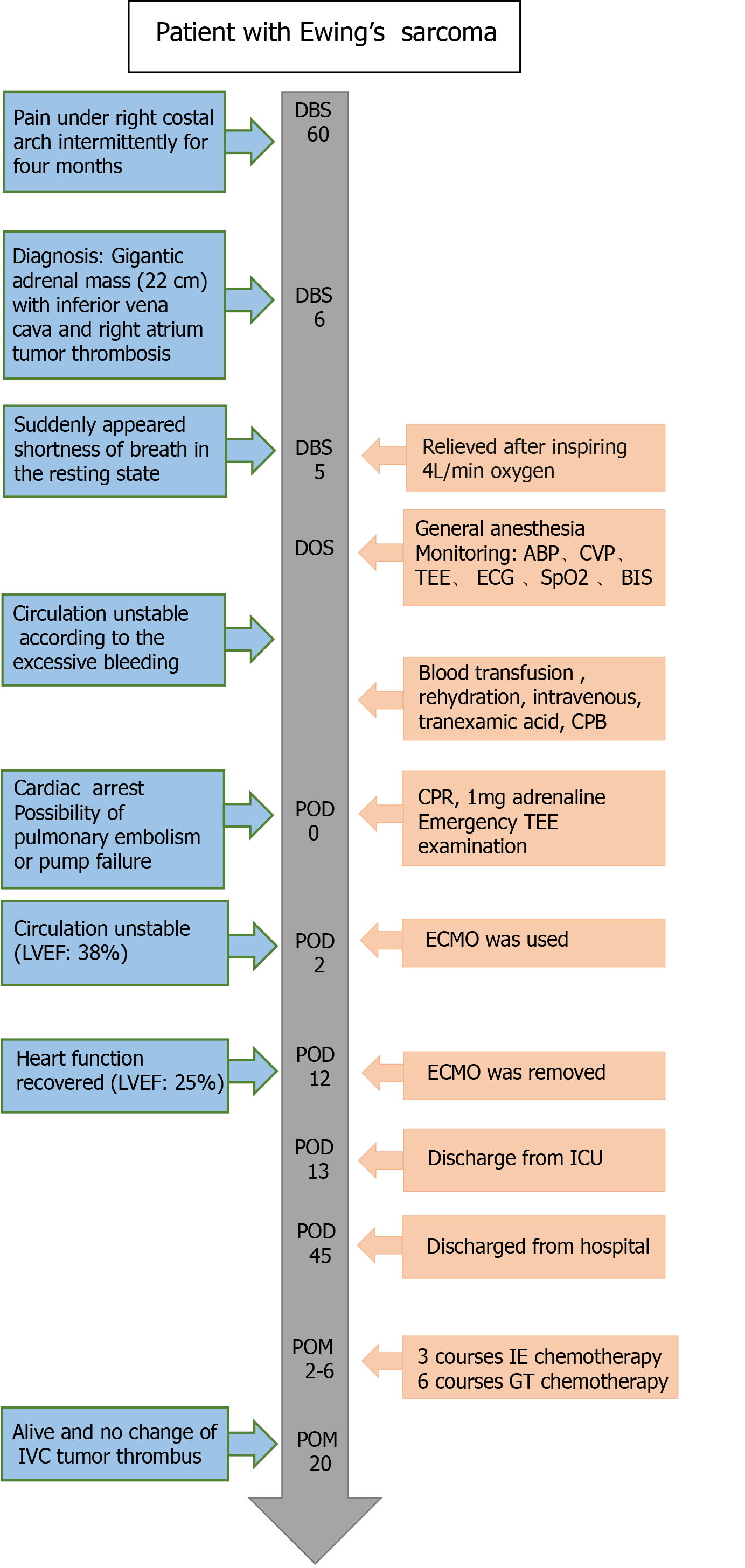
Figure 5 Timeline of perioperative situation, therapies and outcome.
DBS: Day before surgery; DOS: Day of surgery; POD: Postoperative day; LVEF: Left ventricular ejection fraction; TEE: Transesophageal echocardiography; ABP: Arterial blood pressure; CVP: Central venous pressure; ECG: Electrocardiogram; BIS: Bispectral index; CPB: Cardiopulmonary bypass; CPR: Cardiopulmonary resuscitation; TTE: Transthoracic echocardiography; ECMO: Extracorporeal membrane oxygenation; ICU: Intensive care unit.
- Citation: Wang JL, Xu CY, Geng CJ, Liu L, Zhang MZ, Wang H, Xiao RT, Liu L, Zhang G, Ni C, Guo XY. Anesthesia and perioperative management for giant adrenal Ewing’s sarcoma with inferior vena cava and right atrium tumor thrombus: A case report. World J Clin Cases 2022; 10(2): 643-655
- URL: https://www.wjgnet.com/2307-8960/full/v10/i2/643.htm
- DOI: https://dx.doi.org/10.12998/wjcc.v10.i2.643


