Copyright
©The Author(s) 2022.
World J Clin Cases. Jan 14, 2022; 10(2): 631-642
Published online Jan 14, 2022. doi: 10.12998/wjcc.v10.i2.631
Published online Jan 14, 2022. doi: 10.12998/wjcc.v10.i2.631
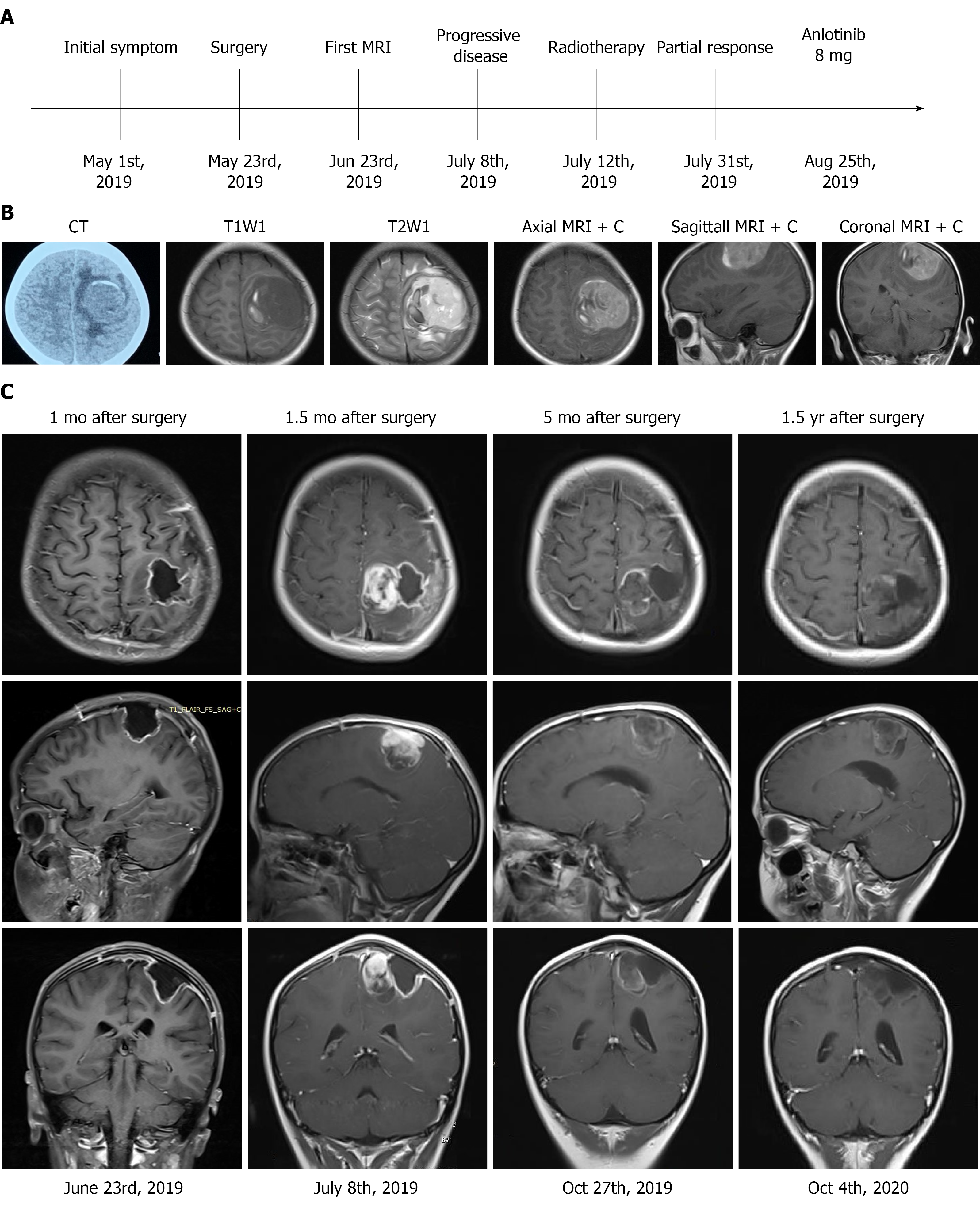
Figure 1 Treatment timeline and radiography.
A: The treatment timeline; B: A head computed tomography scan revealed a quasi-circular mass in the left frontal-parietal region with high-density and associated hemorrhage. Brain magnetic resonance imaging (MRI) revealed low signals on T1 weighted imaging with high surrounding signals. High signals on T2 weighted imaging with low surrounding signals were observed, with marked enhancement on contrast measuring 4.8 cm × 5.0 cm × 4.5 cm in the left motor area of the frontal-parietal lobes (axial, sagittal and coronal views); C: Gadolinium-enhanced MRI imaging in axial, sagittal and coronal views 1 mo after surgery. MRI imaging showed no residual tumor; 1.5 mo after surgery, MRI imaging showed a solid mass. Five months and 1.5 year after surgery, MRI imaging showed that the tumor had not progressed. MRI + C: Gadolinium-enhanced magnetic resonance imaging.
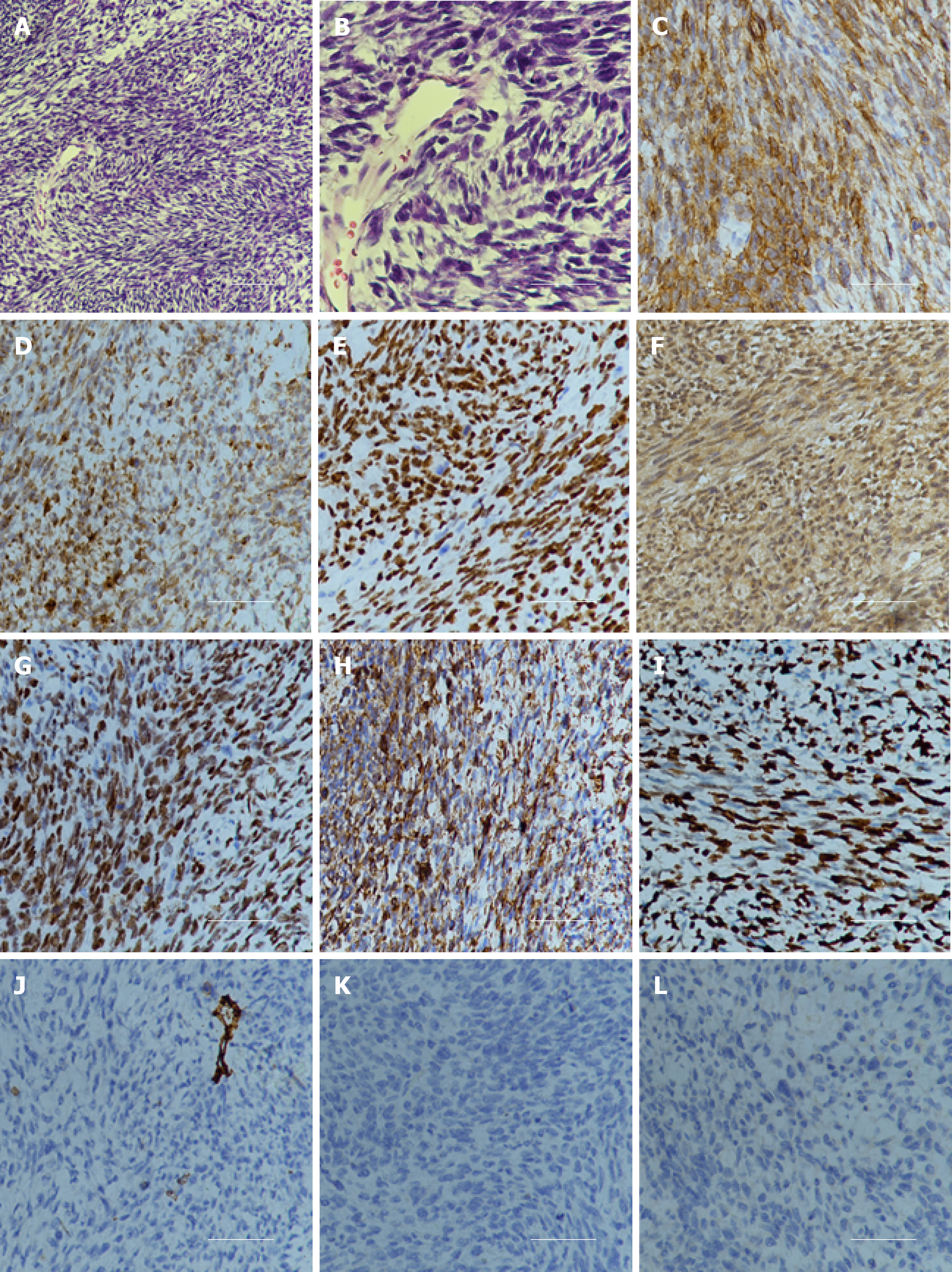
Figure 2 Hematoxylin and eosin staining and immunohistochemistry examination of the specimen.
A: Hematoxylin and eosin staining showed that a large number of spindle or oval cells were diffusely distributed, with deep staining of a null, “staghorn” vascular pattern, hypercellularity and increased mitotic activity were observed in the tumor (> 4 mitosis/10 high-power fields). Hematoxylin and eosin, 100 ×; B: Hematoxylin and eosin staining, 400 ×; C: Positive for CD99, 200 ×; D: Positive for Bcl-2, 200 ×; E: Positive for TP53, 200 ×; F: Positive for IDH1, 200 ×; G: Positive for TLE-1, 200 ×; H: Positive for vimentin, 200 ×; I: High Ki-67 proliferation index: 80%, 200 ×; J: Negative for CD34, 200 ×; K: Negative for STAT6, 200 ×; L: Negative for S100, 200 ×.
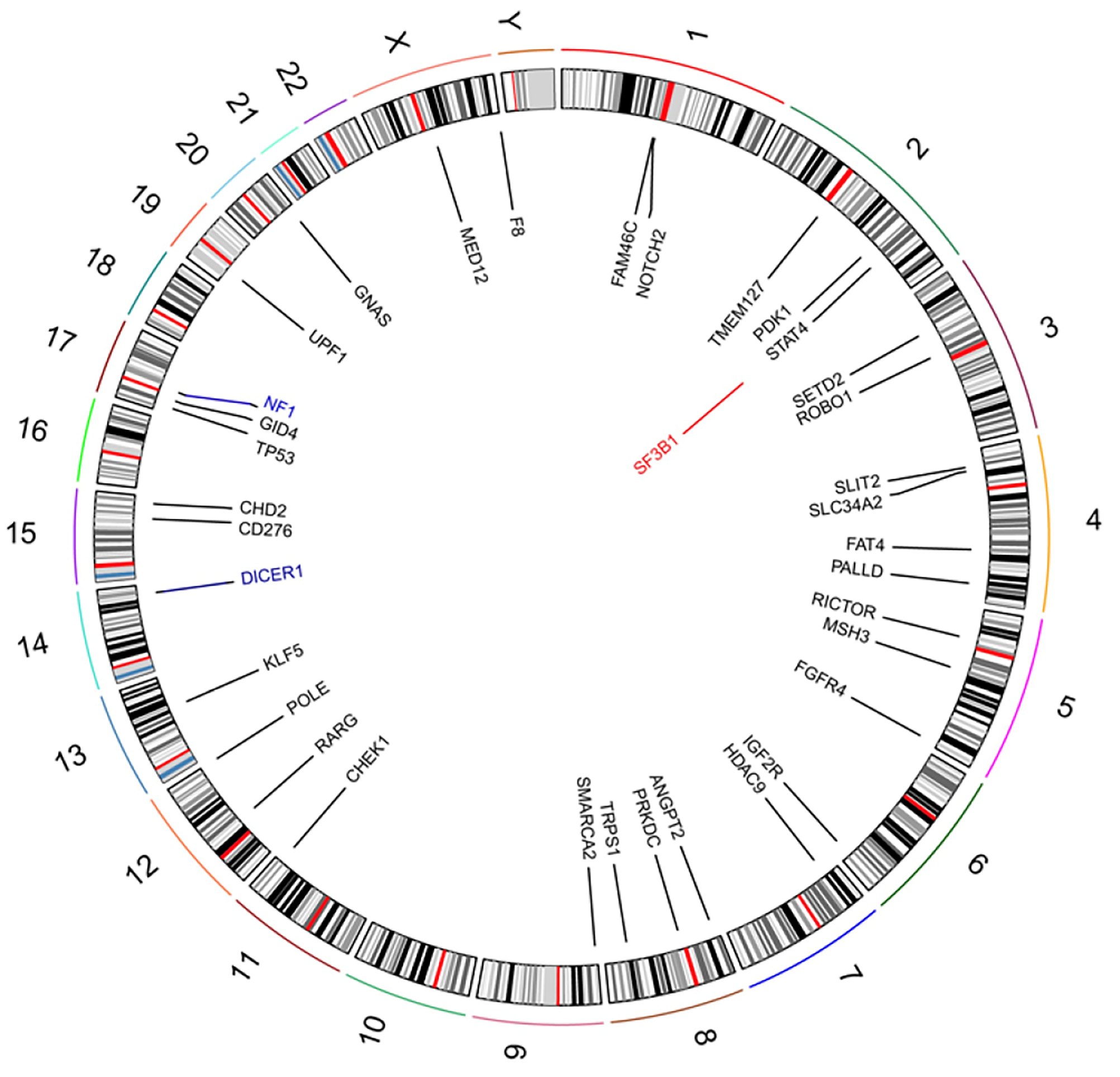
Figure 3 Circos 2D track plot of somatic variant across chromosomes in solitary fibrous tumor.
In the inner ring, black denotes missense mutation, blue denotes frameshift mutation, and red denotes gene amplification.
- Citation: Zhang DY, Su L, Wang YW. Malignant solitary fibrous tumor in the central nervous system treated with surgery, radiotherapy and anlotinib: A case report. World J Clin Cases 2022; 10(2): 631-642
- URL: https://www.wjgnet.com/2307-8960/full/v10/i2/631.htm
- DOI: https://dx.doi.org/10.12998/wjcc.v10.i2.631


