Copyright
©The Author(s) 2022.
World J Clin Cases. Jan 14, 2022; 10(2): 618-624
Published online Jan 14, 2022. doi: 10.12998/wjcc.v10.i2.618
Published online Jan 14, 2022. doi: 10.12998/wjcc.v10.i2.618
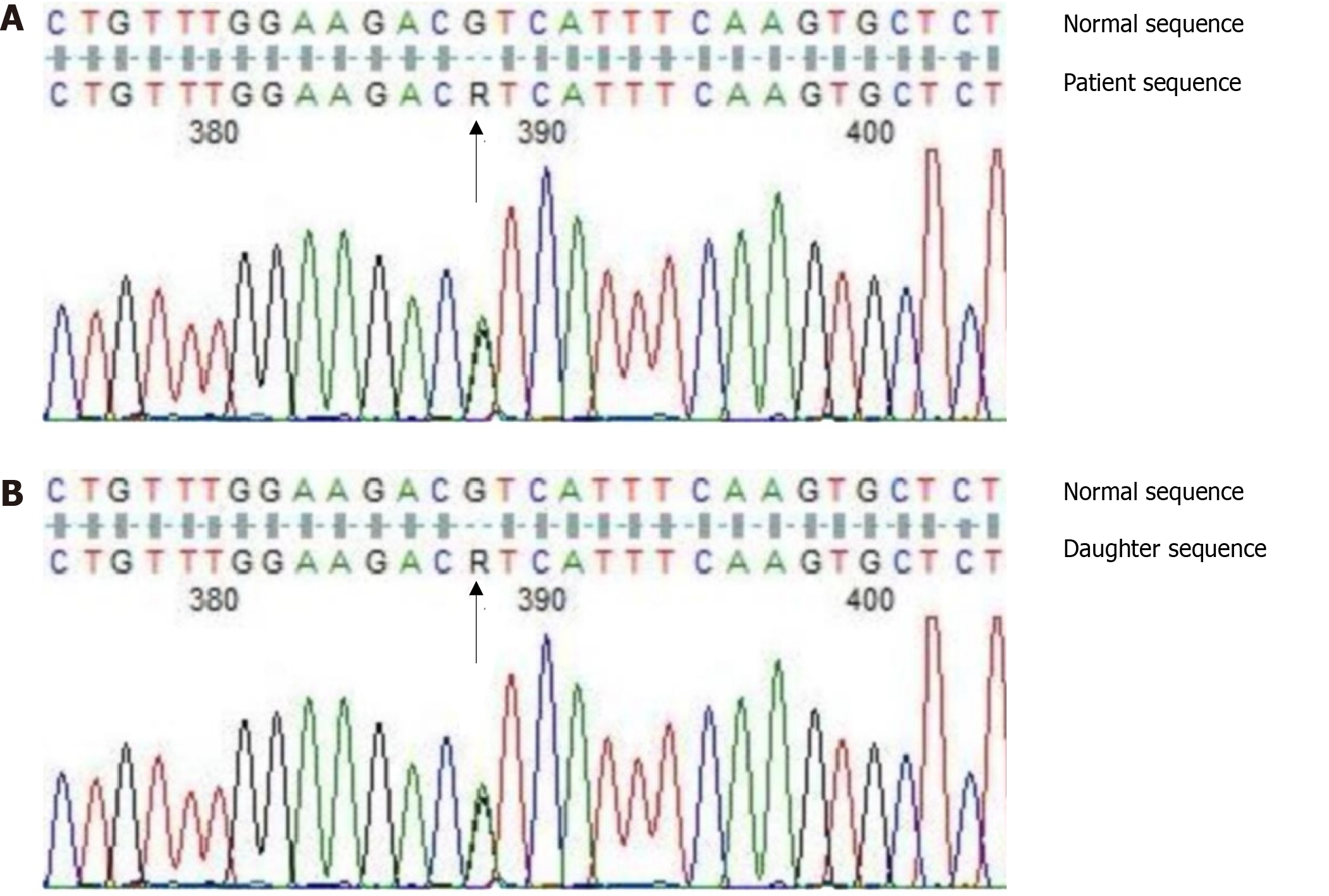
Figure 1 Sequence map illustrating the SERPINC1 gene mutation in the patient (A) and his daughter (B).
The heterozygous mutation site (rs2227589 polymorphism site) of c.41+141G>A is marked by the black arrow.
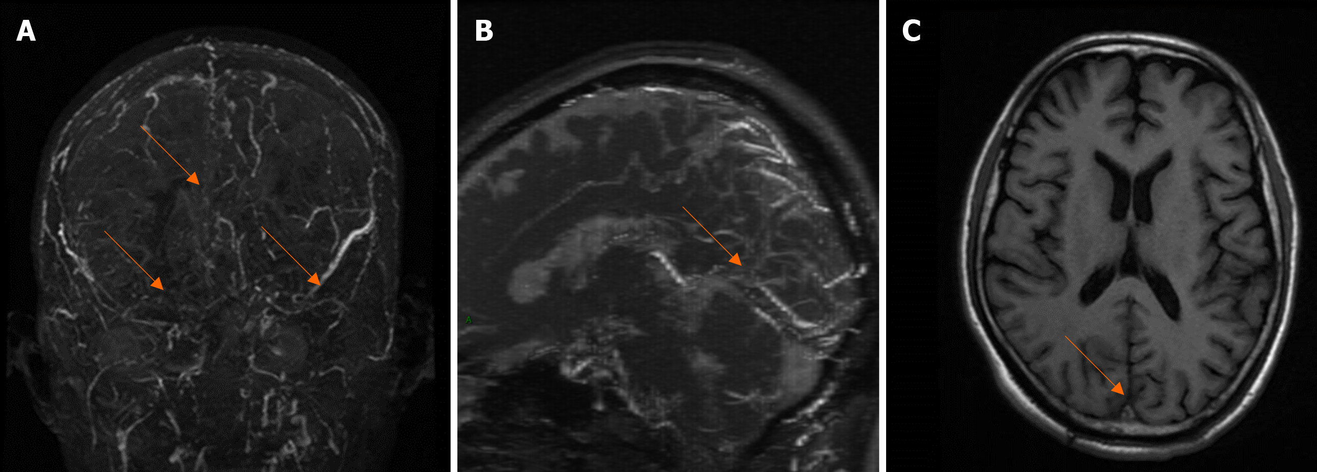
Figure 2 Brain magnetic resonance imaging at admission.
A: Filling defects seen in the superior sagittal sinus, bilateral transverse sinuses, and sigmoid sinus, indicated by magnetic resonance venography (MRV) are shown by arrows; B: Blurred image of the inferior sagittal sinus and straight sinus, indicated by MRV are shown by arrows; C: Thrombosis of the superior sagittal sinus, indicated by magnetic resonance imaging is shown by the arrow.
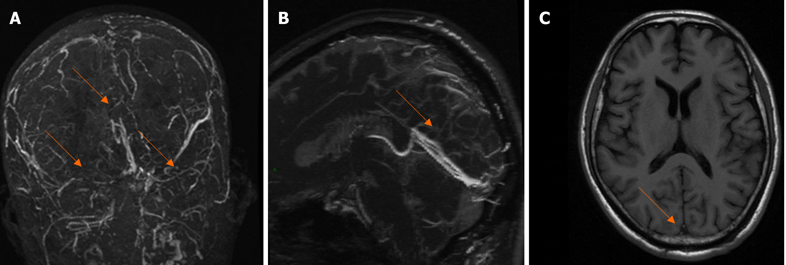
Figure 3 Brain magnetic resonance imaging taken 2 wk after treatment.
A: Bilateral superior sagittal sinuses, bilateral transverse sinus and sigmoid sinus enlarged with respect to Figure 1, indicated by magnetic resonance venography (MRV) are shown by arrows; B: Inferior sagittal sinus and straight sinus are significantly less occluded with respect to Figure 1, indicated by MRV as shown by the arrow; C: Thrombosis shadow seen in the superior sagittal sinus is reduced in size with respect to Figure 1, indicated by arrow.
- Citation: Liao F, Zeng JL, Pan JG, Ma J, Zhang ZJ, Lin ZJ, Lin LF, Chen YS, Ma XT. Patients with SERPINC1 rs2227589 polymorphism found to have multiple cerebral venous sinus thromboses despite a normal antithrombin level: A case report. World J Clin Cases 2022; 10(2): 618-624
- URL: https://www.wjgnet.com/2307-8960/full/v10/i2/618.htm
- DOI: https://dx.doi.org/10.12998/wjcc.v10.i2.618


