Copyright
©The Author(s) 2022.
World J Clin Cases. Jan 14, 2022; 10(2): 528-537
Published online Jan 14, 2022. doi: 10.12998/wjcc.v10.i2.528
Published online Jan 14, 2022. doi: 10.12998/wjcc.v10.i2.528
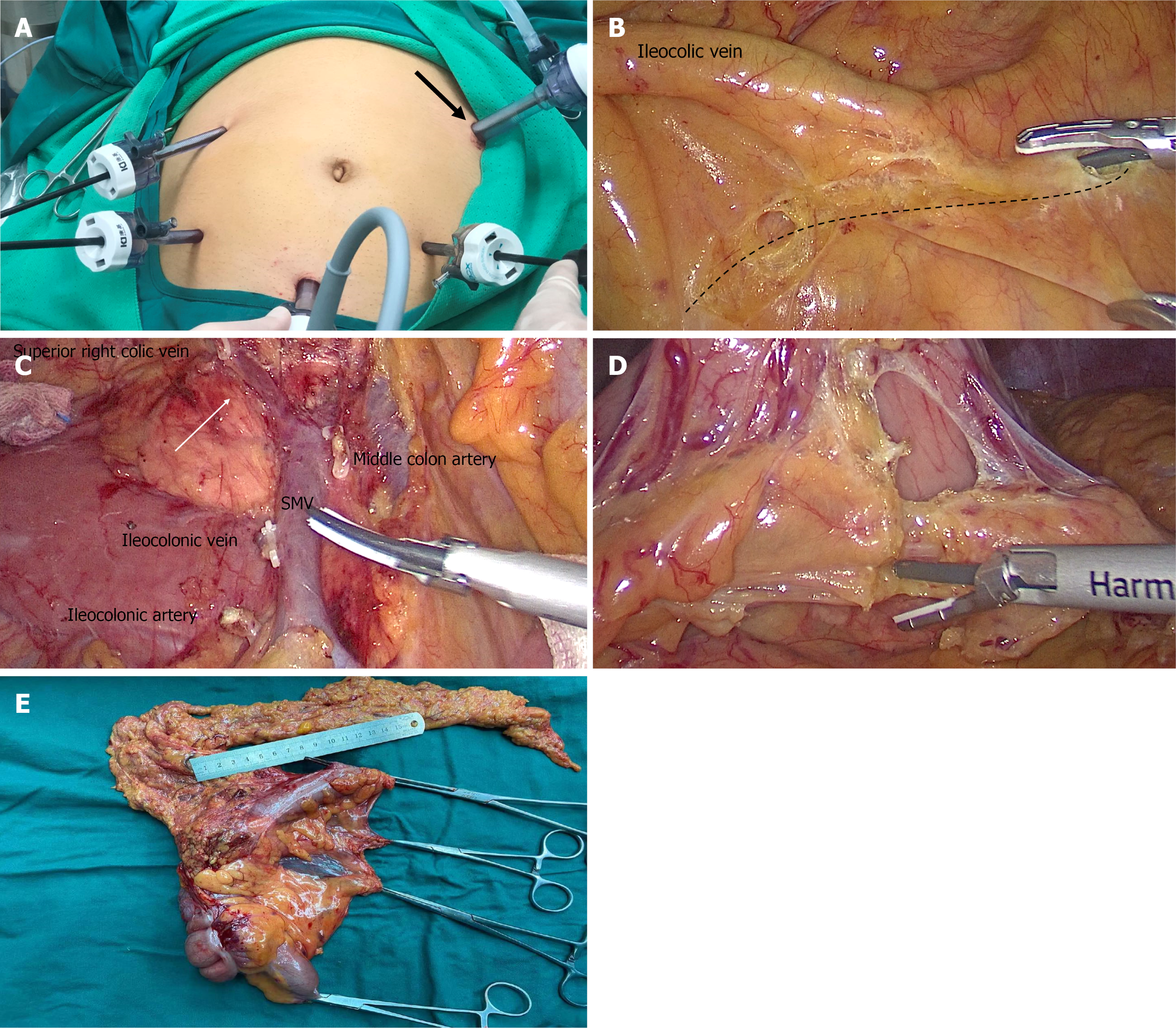
Figure 1 Surgery process photos.
A: Five-hole method and main operation hole (black arrow); B: Caudal ventral approach in which the assistant lifts the ileocolonic vascular pedicle, and an ultrasonic knife is inserted obliquely in the direction of the superior mesenteric vein (SMV) (dotted line) into the small intestine ascending colon space; C: SMV with broken branches and Henle trunk (white arrow); D: Dissection of gastrocolic ligament lymph nodes; (E) Postoperative specimens.
- Citation: Zheng HD, Xu JH, Liu YR, Sun YF. Analysis of 20 patients with laparoscopic extended right colectomy. World J Clin Cases 2022; 10(2): 528-537
- URL: https://www.wjgnet.com/2307-8960/full/v10/i2/528.htm
- DOI: https://dx.doi.org/10.12998/wjcc.v10.i2.528


