Copyright
©The Author(s) 2022.
World J Clin Cases. Jan 14, 2022; 10(2): 437-447
Published online Jan 14, 2022. doi: 10.12998/wjcc.v10.i2.437
Published online Jan 14, 2022. doi: 10.12998/wjcc.v10.i2.437
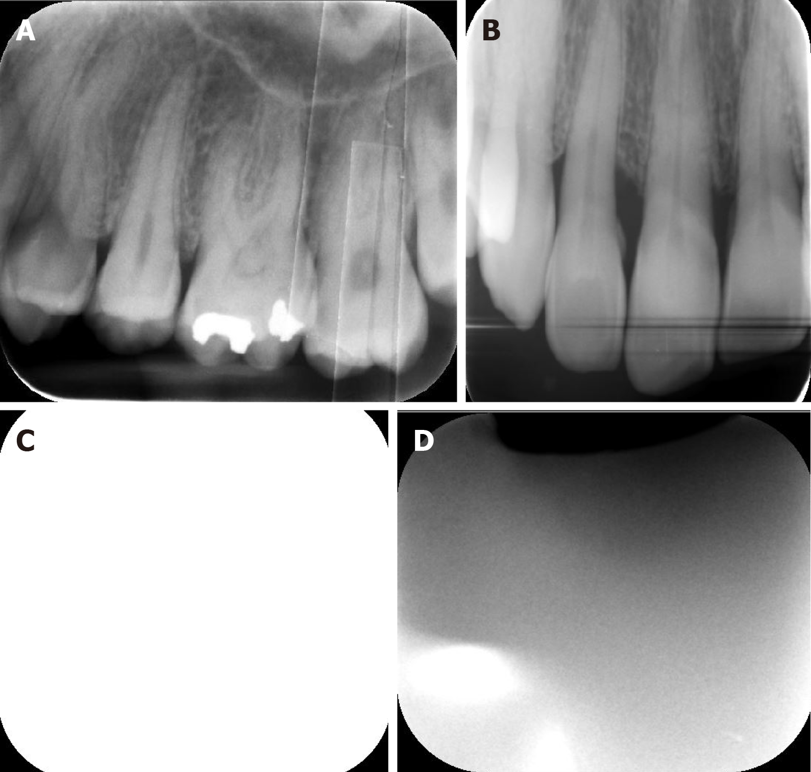
Figure 1 Periapical and bitewing radiographs show some operator errors.
A: Movement of phosphor plate inside the disposable packet; B: Movement of phosphor plate in the disposable packet and plate scratch; C: Reversed image, cone cut and white line; D: Reversed image and improper plate placement.
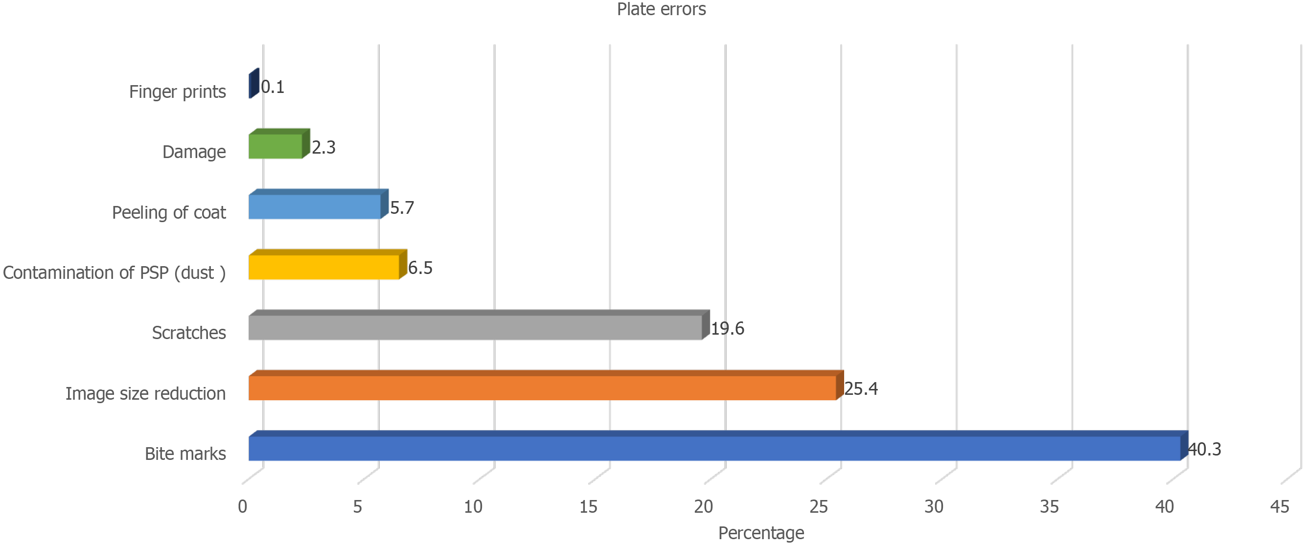
Figure 2 Bar chart illustrating frequency and percentage of plate errors.
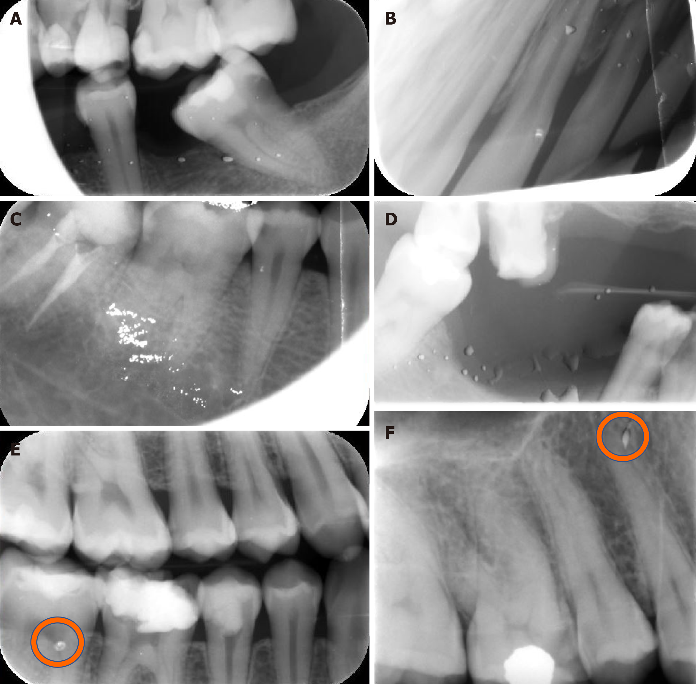
Figure 3 Bitewing and periapical radiographs show some plate errors.
A: Plate contamination and cone cut; B: Plate contamination, plate movement inside packet, white line, black line, and image distortion; C: Plate contamination, white line and cone cut; D: Plate contamination and non-uniform image; E: Bitemarks; F: Plate scratch.
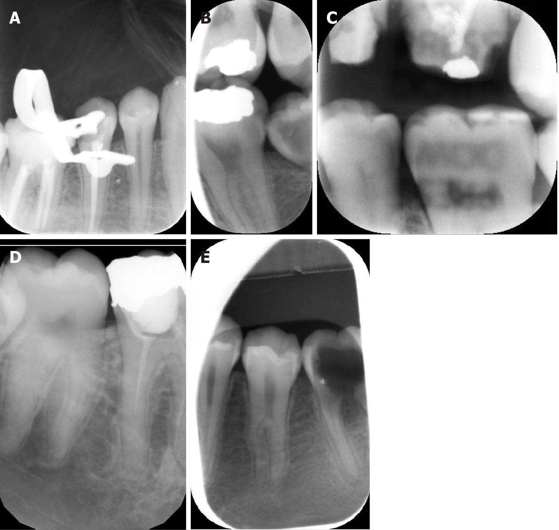
Figure 4 Periapical and bitewing radiographs show some plate errors.
A: Plate scratches; B-D: image size reduction; E: Image size reduction and movement of plate inside packet.
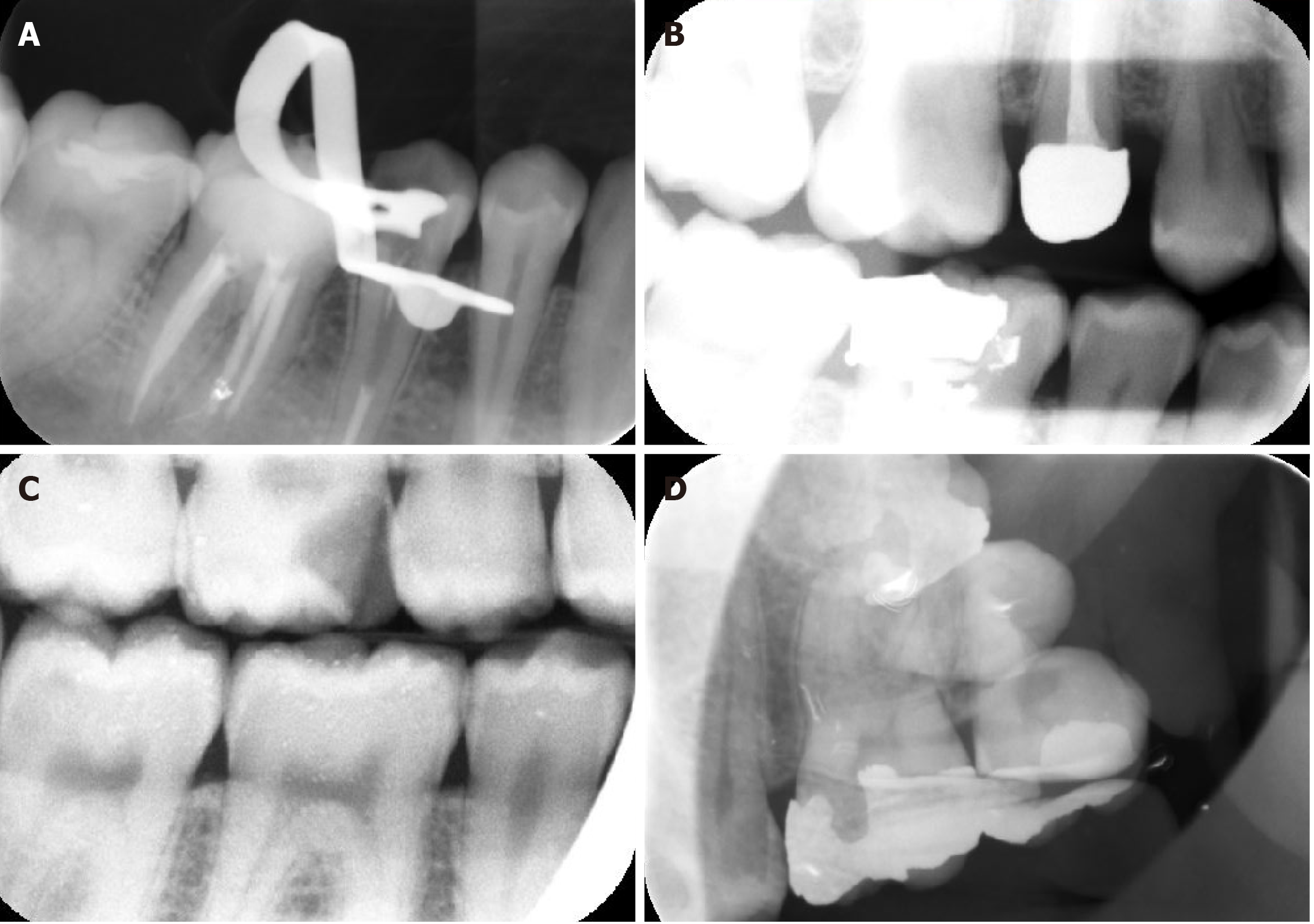
Figure 5 Periapical and bitewing radiographs show some scanning errors.
A and B: Non-uniformity of the image; C: bright noisy image; D: Double image due to incomplete erasing.
Figure 6 Periapical radiographs show some scanning errors.
A: Multiple white lines; B: Black line: C: Blank image; D: Inhomogeneous image.
- Citation: Elkhateeb SM, Aloyouny AY, Omer MMS, Mansour SM. Analysis of photostimulable phosphor image plate artifacts and their prevalence. World J Clin Cases 2022; 10(2): 437-447
- URL: https://www.wjgnet.com/2307-8960/full/v10/i2/437.htm
- DOI: https://dx.doi.org/10.12998/wjcc.v10.i2.437


