Copyright
©The Author(s) 2022.
World J Clin Cases. Jul 6, 2022; 10(19): 6722-6727
Published online Jul 6, 2022. doi: 10.12998/wjcc.v10.i19.6722
Published online Jul 6, 2022. doi: 10.12998/wjcc.v10.i19.6722
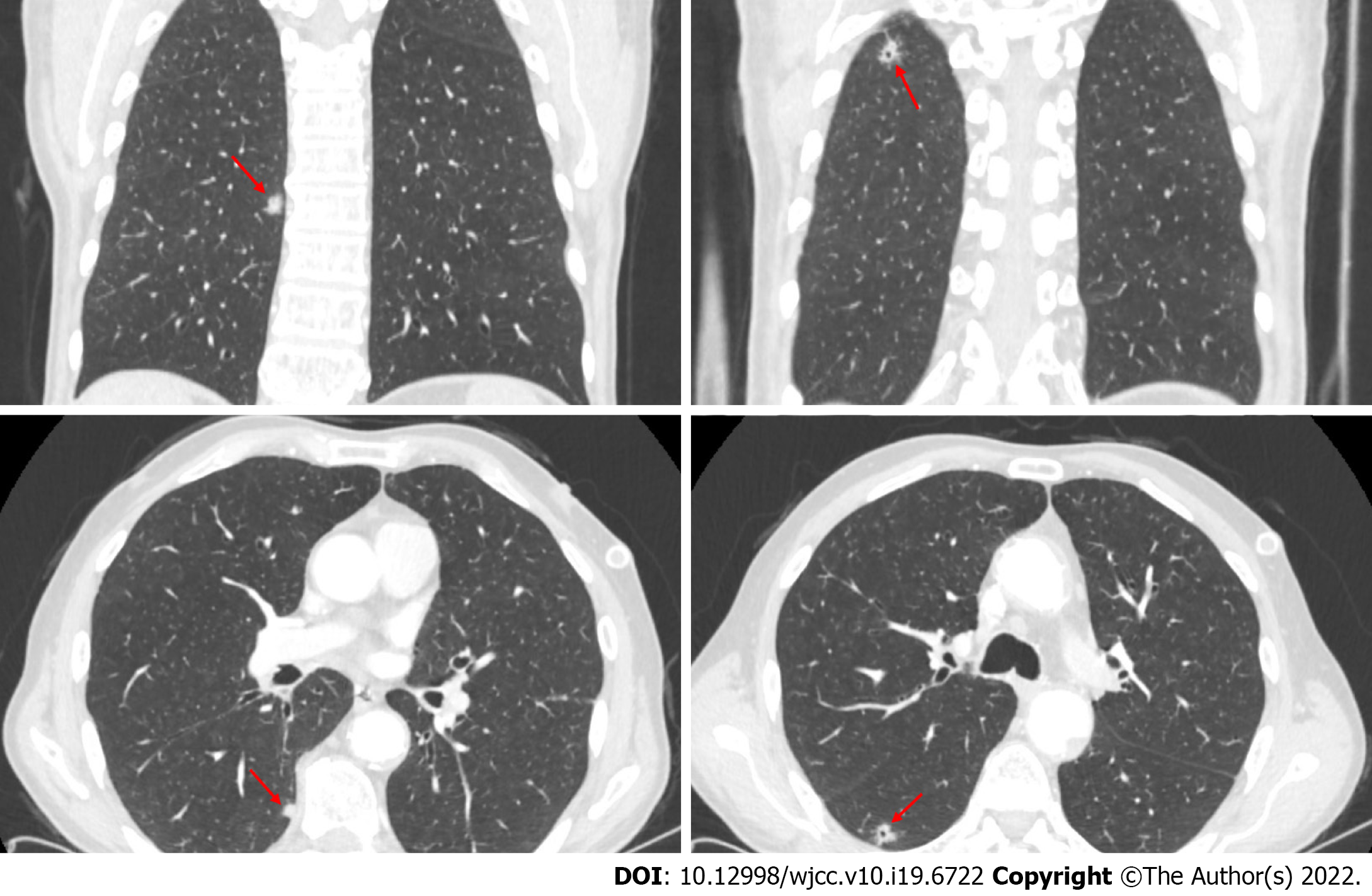
Figure 1 Location of nodules on computed tomography images.
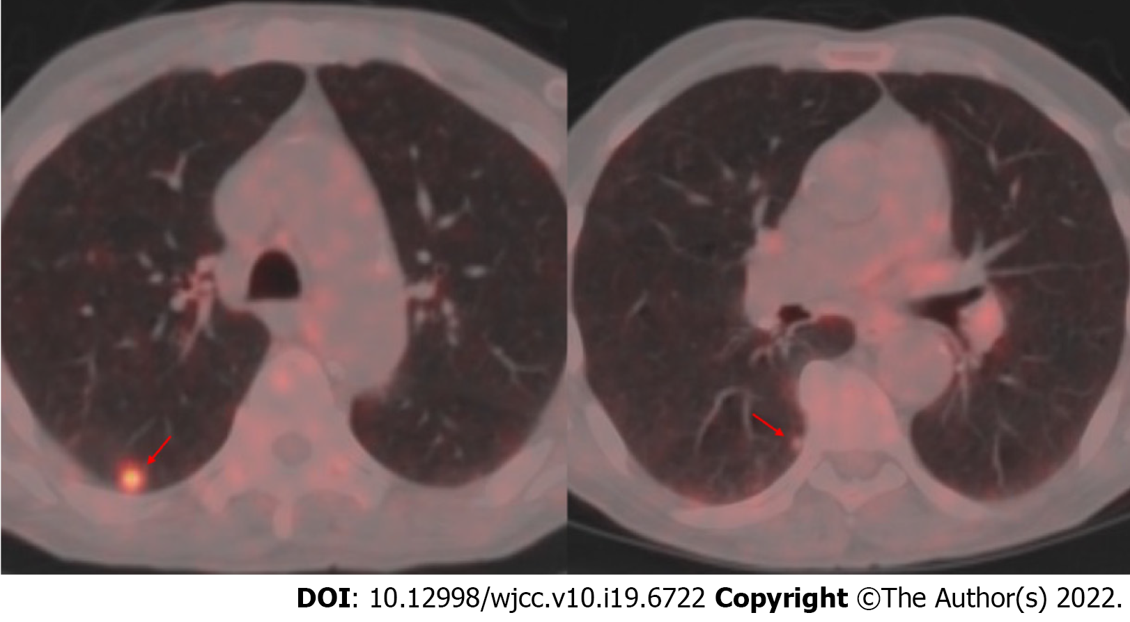
Figure 2 Fludeoxyglucose uptake in nodules in Positron emission tomography - computed tomography.
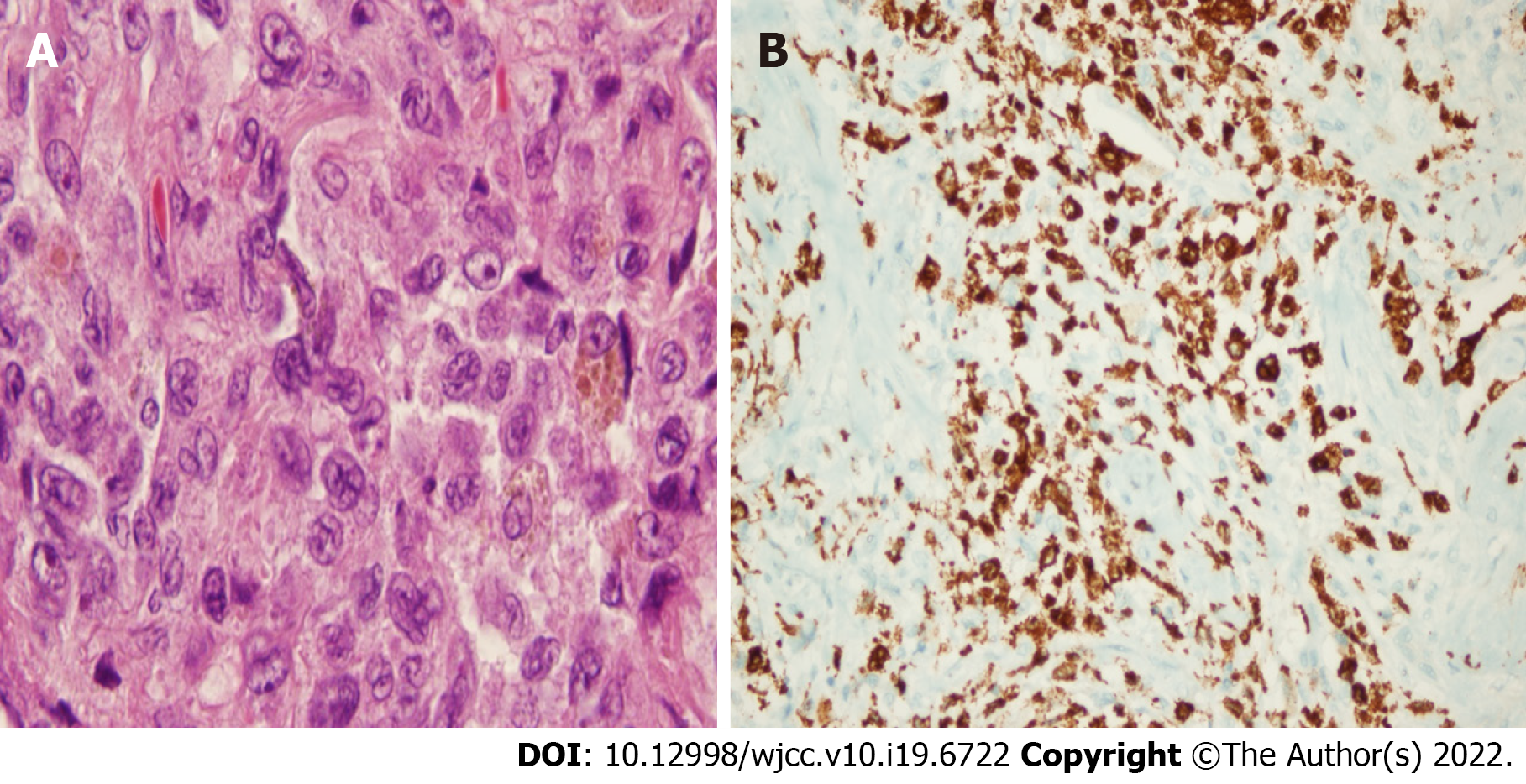
Figure 3 Immunohistochemical analyses in Langerhans cell histiocytosis.
A: Characteristic Langerhans cells in a nodule with pulmonary Langerhans cell histiocytosis (hematoxylin-eosin, × 100); B: Histopathologic diagnosis of Pulmonary Langerhans cell histiocytosis from tissue blocks was supported by immunohistochemistry staining analyses for Langerin.
- Citation: Gencer A, Ozcibik G, Karakas FG, Sarbay I, Batur S, Borekci S, Turna A. Two smoking-related lesions in the same pulmonary lobe of squamous cell carcinoma and pulmonary Langerhans cell histiocytosis: A case report . World J Clin Cases 2022; 10(19): 6722-6727
- URL: https://www.wjgnet.com/2307-8960/full/v10/i19/6722.htm
- DOI: https://dx.doi.org/10.12998/wjcc.v10.i19.6722


