Copyright
©The Author(s) 2022.
World J Clin Cases. Jul 6, 2022; 10(19): 6602-6608
Published online Jul 6, 2022. doi: 10.12998/wjcc.v10.i19.6602
Published online Jul 6, 2022. doi: 10.12998/wjcc.v10.i19.6602
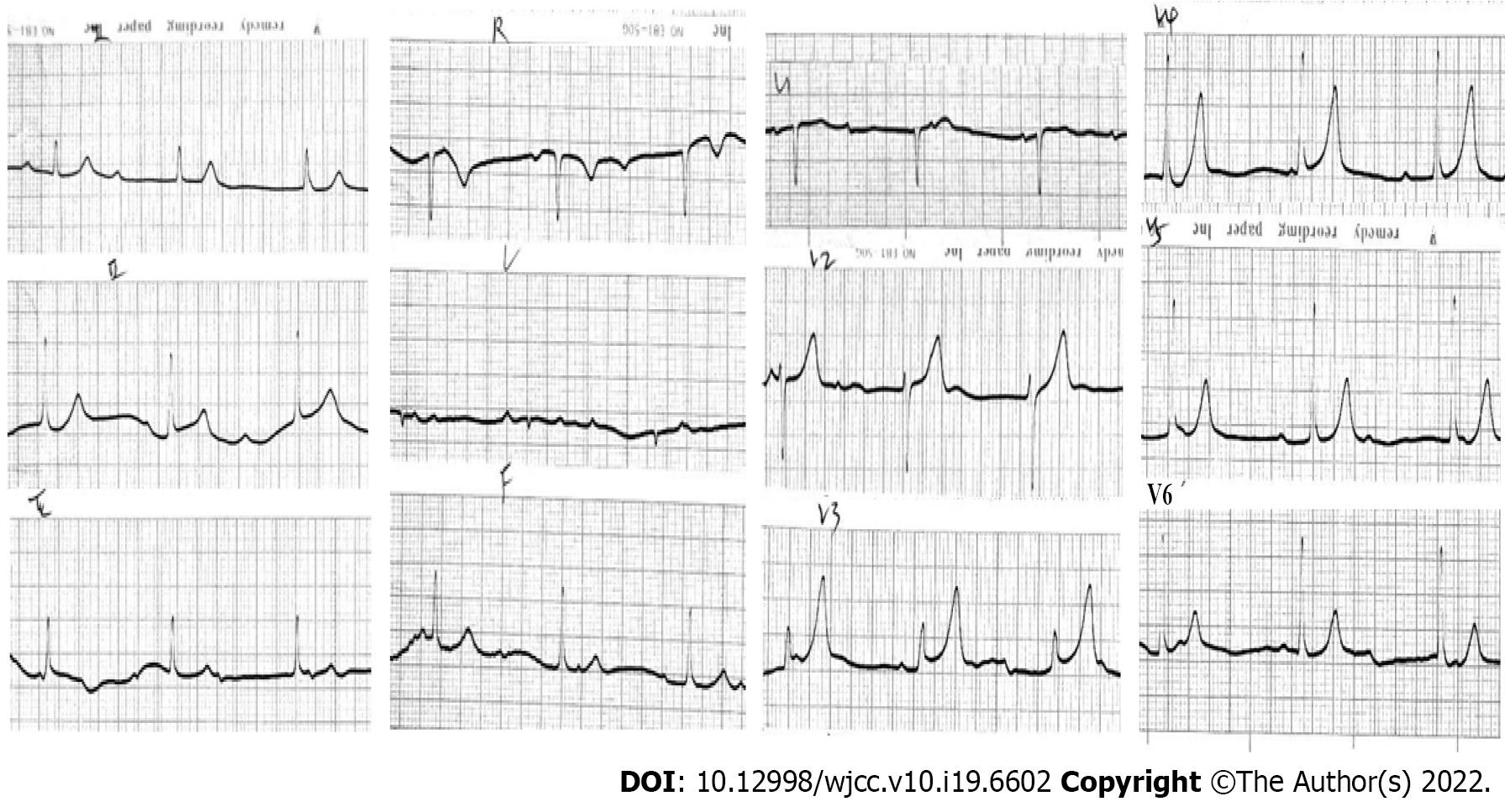
Figure 1 Electrocardiography on admission on admission.
Complete atrioventricular block with junctional escape at 48 beats per minute.
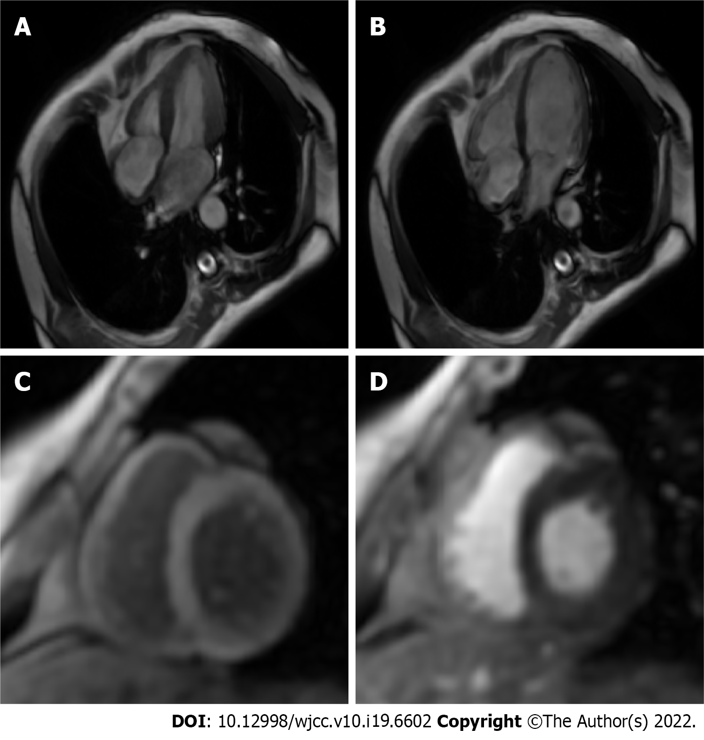
Figure 2 Cardiovascular contrast-enhanced magnetic resonance imaging.
A: Cardiac magnetic resonance image (MRI) of the systolic phase of four chambers and axial view showed normal atrioventricular size and myocardial function; B: Cardiac MRI of the diastolic phase of four chambers and axial view showed normal atrioventricular size and myocardial function; C: T2-weighted image did not reveal any myocardial edema; D: Late gadolinium enhancement revealed no definite area of hyperenhancement to suggest myocardial fibrosis.
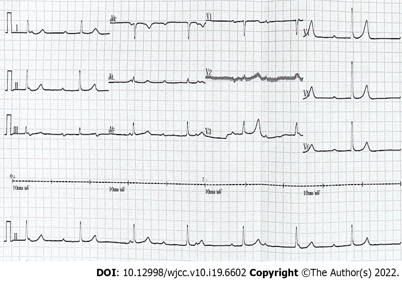
Figure 3 Electrocardiography at 28 years of follow-up.
Complete atrioventricular block with junctional escape at 42 beats per minute.
- Citation: Su LN, Wu MY, Cui YX, Lee CY, Song JX, Chen H. Unusual course of congenital complete heart block in an adult: A case report. World J Clin Cases 2022; 10(19): 6602-6608
- URL: https://www.wjgnet.com/2307-8960/full/v10/i19/6602.htm
- DOI: https://dx.doi.org/10.12998/wjcc.v10.i19.6602


