Copyright
©The Author(s) 2022.
World J Clin Cases. Jul 6, 2022; 10(19): 6587-6594
Published online Jul 6, 2022. doi: 10.12998/wjcc.v10.i19.6587
Published online Jul 6, 2022. doi: 10.12998/wjcc.v10.i19.6587
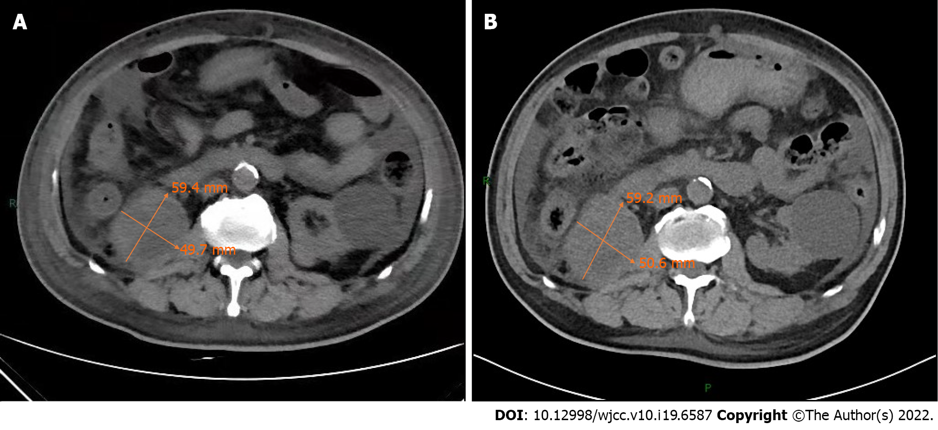
Figure 1 Plain abdomen computed tomography scan.
A: Plain abdomen computed tomography scan at admission showed an enlarged right kidney, irregular soft tissue occupying, unclear boundary, and an about 59.4 mm× 49.7 mm larger layer. Compared with previous computed tomography, there was no significant change in tumor size; B: Plain abdomen computed tomography scan on February 2, 2021 showed an enlarged right kidney, irregular soft tissue occupying, unclear boundary, and an about 59.2 mm × 50.6 mm larger layer.
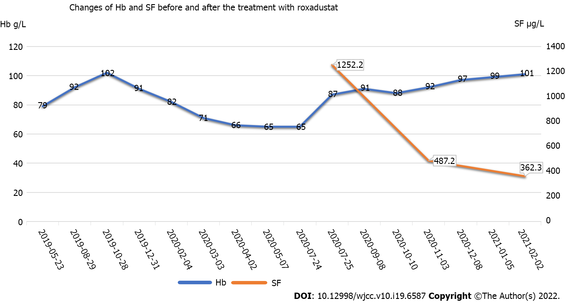
Figure 2 Dynamic changes of hemoglobin and serum ferritin.
The patient was admitted to our hospital on July 24, 2020. He was treated with 1.5 units of red blood cell transfusion on that day. After discharge, he began to take 20 mg oral roxadustat three times a week on August 1, 2020. Then he began to take 50 mg oral roxadustat three times a week on December 8, 2020. Hb: Hemoglobin; SF: Serum ferritin.
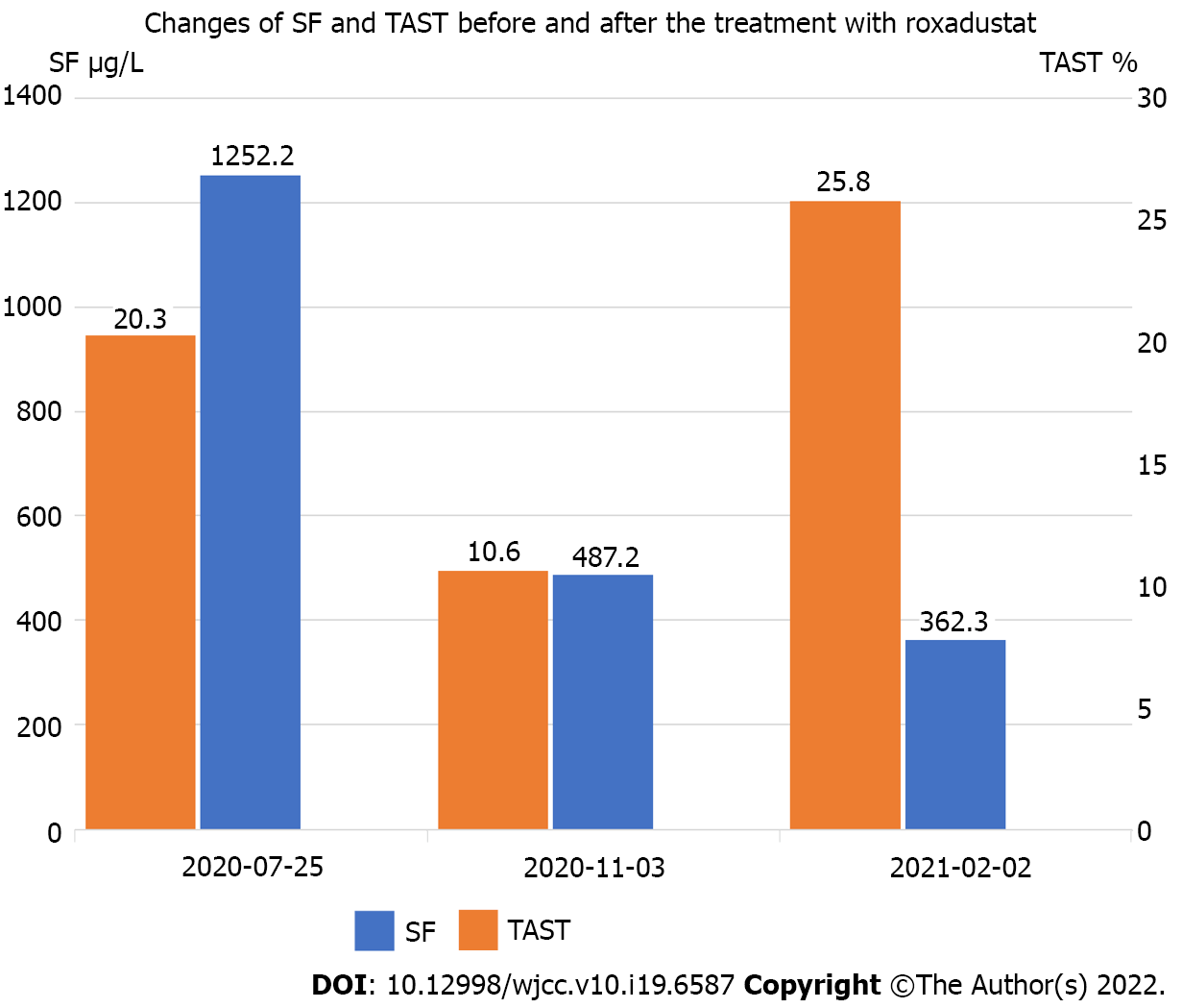
Figure 3 Changes of serum ferritin and transferrin saturation.
The patient’s serum ferritin was as high as 1252.2 mg/L on the second day after blood transfusion on July 25, 2020. His iron index was further improved after taking roxadustat orally in the following months. SF: Serum ferritin; TAST: Transferrin saturation.
- Citation: Zhou QQ, Li J, Liu B, Wang CL. Roxadustat for treatment of anemia in a cancer patient with end-stage renal disease: A case report. World J Clin Cases 2022; 10(19): 6587-6594
- URL: https://www.wjgnet.com/2307-8960/full/v10/i19/6587.htm
- DOI: https://dx.doi.org/10.12998/wjcc.v10.i19.6587


