Copyright
©The Author(s) 2022.
World J Clin Cases. Jul 6, 2022; 10(19): 6507-6513
Published online Jul 6, 2022. doi: 10.12998/wjcc.v10.i19.6507
Published online Jul 6, 2022. doi: 10.12998/wjcc.v10.i19.6507
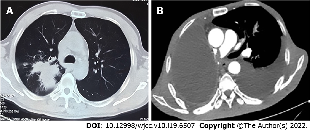
Figure 1 Chest computed tomography and enhanced computed tomography.
A: Computed tomography (CT) revealed a mass at the right hilum prior to performing radical resection of right lung cancer; B: Enhanced CT revealed pulmonary embolism after bevacizumab combined with chemotherapy therapy.
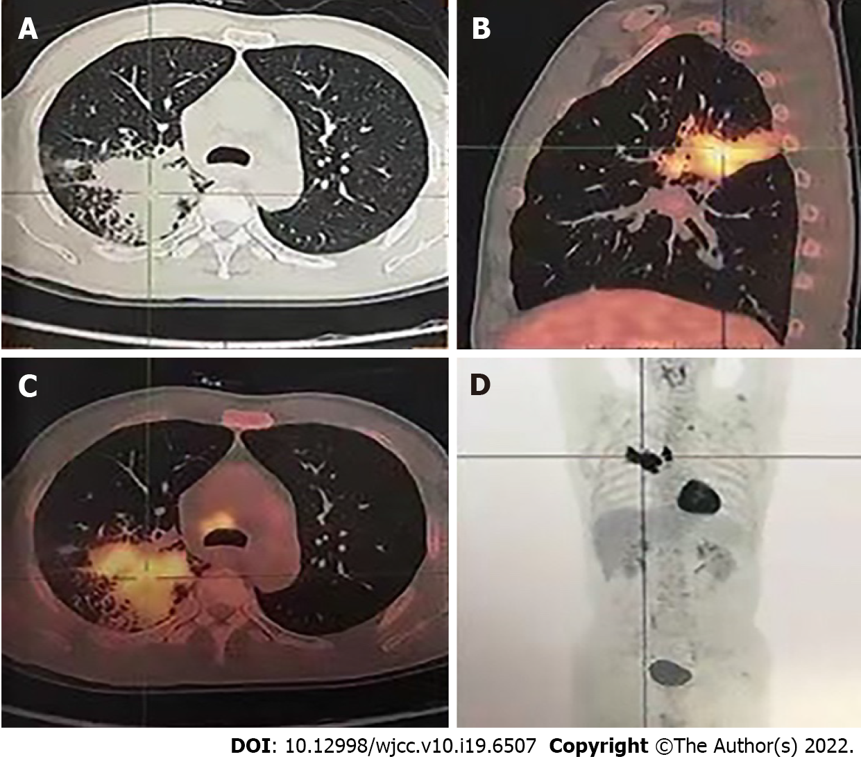
Figure 2 Positron emission tomography-computed tomography before radical resection of right lung cancer.
A: Irregular soft tissue mass in the right lung hilum; B: Posterior wall cavity with abnormal concentration of radioactivity in the mass; C: Consolidation and thick-walled voids in the posterior and dorsal lobes of the right lung; D: Mediastinal lymph node metastasis with no distant metastases.
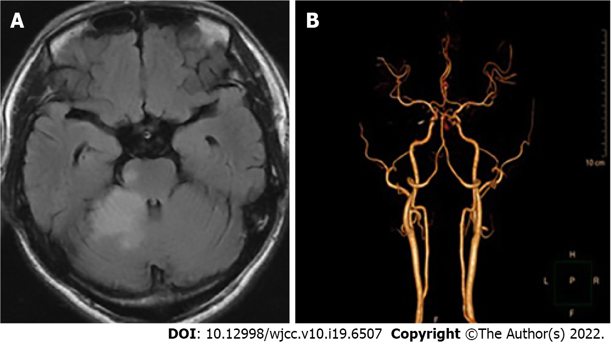
Figure 3 Magnetic resonance imaging and computed tomography angiography of the head.
A: Magnetic resonance imaging revealed cerebral infarction following treatment using bevacizumab combined with chemotherapy; B: Computed tomography angiography revealed local occlusion of the right superior cerebellar artery.
- Citation: Kong Y, Xu XC, Hong L. Arteriovenous thrombotic events in a patient with advanced lung cancer following bevacizumab plus chemotherapy: A case report. World J Clin Cases 2022; 10(19): 6507-6513
- URL: https://www.wjgnet.com/2307-8960/full/v10/i19/6507.htm
- DOI: https://dx.doi.org/10.12998/wjcc.v10.i19.6507


