Copyright
©The Author(s) 2022.
World J Clin Cases. Jun 26, 2022; 10(18): 6333-6337
Published online Jun 26, 2022. doi: 10.12998/wjcc.v10.i18.6333
Published online Jun 26, 2022. doi: 10.12998/wjcc.v10.i18.6333
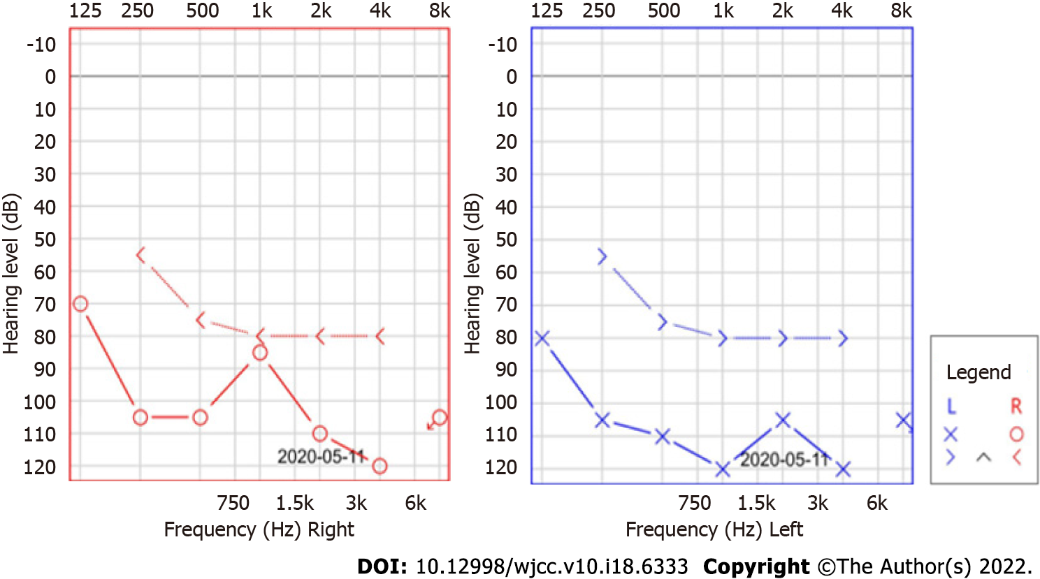
Figure 1 Pure tone audiometry.
Pure tone audiometry revealed severe bilateral low- and high-frequency hearing loss.
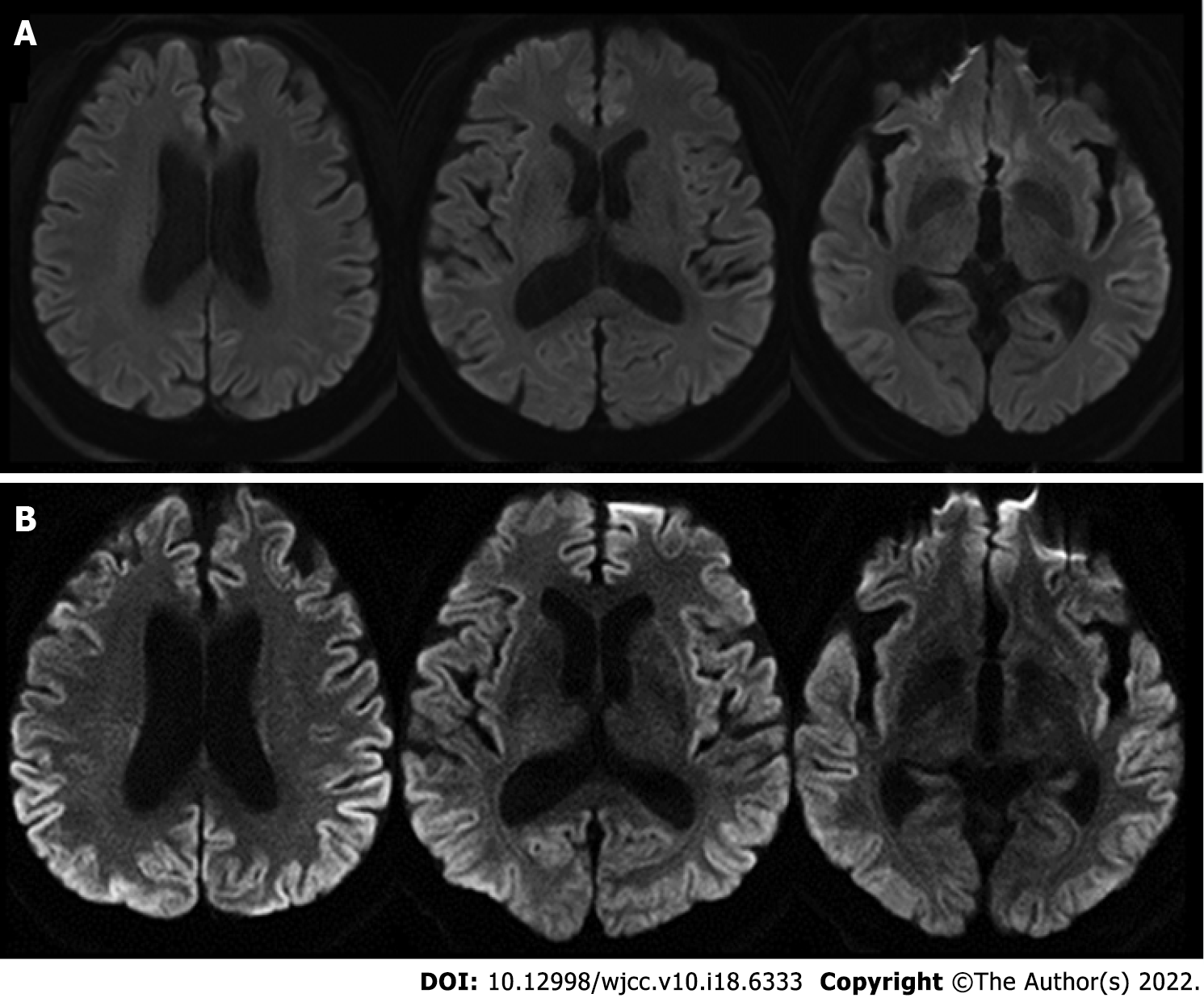
Figure 2 Findings of brain magnetic resonance imaging.
A: The initial brain magnetic resonance imaging (MRI). The initial brain diffusion-weighted imaging was unremarkable; B: The follow-up brain MRI. The follow-up brain diffusion-weighted imaging showed high signal intensities in the bilateral frontal, temporal, parietal, and occipital cortices.
- Citation: Na S, Lee SA, Lee JD, Lee ES, Lee TK. Creutzfeldt-Jakob disease presenting with bilateral hearing loss: A case report. World J Clin Cases 2022; 10(18): 6333-6337
- URL: https://www.wjgnet.com/2307-8960/full/v10/i18/6333.htm
- DOI: https://dx.doi.org/10.12998/wjcc.v10.i18.6333


