Copyright
©The Author(s) 2022.
World J Clin Cases. Jun 26, 2022; 10(18): 6314-6318
Published online Jun 26, 2022. doi: 10.12998/wjcc.v10.i18.6314
Published online Jun 26, 2022. doi: 10.12998/wjcc.v10.i18.6314
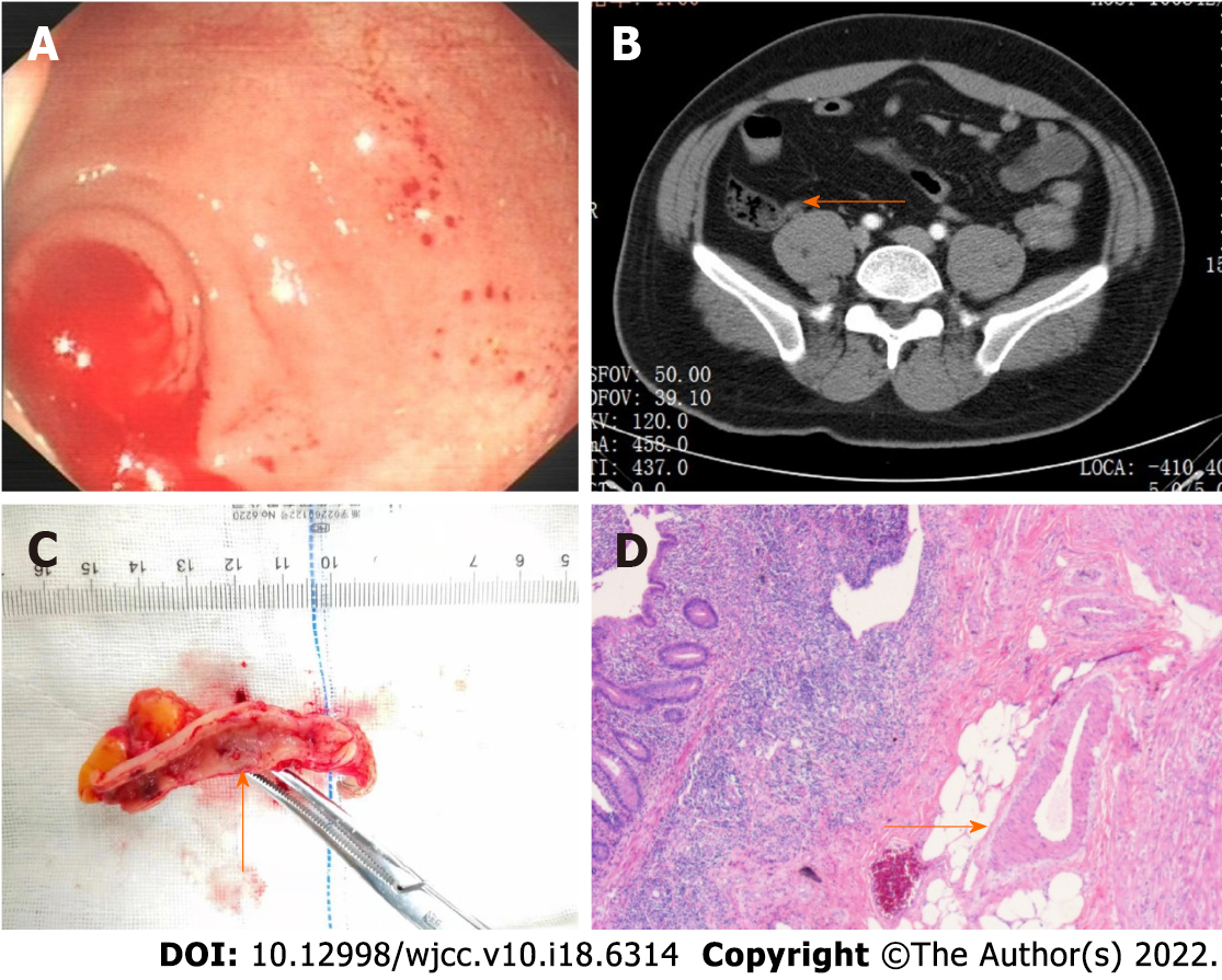
Figure 1 Appendiceal bleeding caused by Dieulafoy’s lesion.
A: Colonoscopy showed active bleeding from the appendiceal orifice after blood clots were flushed out of the ileocecal junction; B: Enhanced abdominal computed tomography showed a high-density area in the appendix, revealing the possibility of appendiceal bleeding (arrow); C: Macroscopic pathological observation showed a vessel stump on the mucosa of the appendix (arrow); D: Microscopic pathological examination showed a caliber-persistent artery in the submucosa of the appendix (arrow).
- Citation: Zhou SY, Guo MD, Ye XH. Appendiceal bleeding: A case report. World J Clin Cases 2022; 10(18): 6314-6318
- URL: https://www.wjgnet.com/2307-8960/full/v10/i18/6314.htm
- DOI: https://dx.doi.org/10.12998/wjcc.v10.i18.6314


