Copyright
©The Author(s) 2022.
World J Clin Cases. Jun 26, 2022; 10(18): 6298-6306
Published online Jun 26, 2022. doi: 10.12998/wjcc.v10.i18.6298
Published online Jun 26, 2022. doi: 10.12998/wjcc.v10.i18.6298
Figure 1 Initial intraoral photographs.
A: Case 1, bleeding from the gingival crevice of tooth 11; B: Case 2, complicated crown fracture in tooth 11 with discolored crown and pulp chamber filled by resin, and mild inflammation in the marginal gingiva of teeth 11 and 21; C: Case 1, tooth 11 taken 1 yr later.
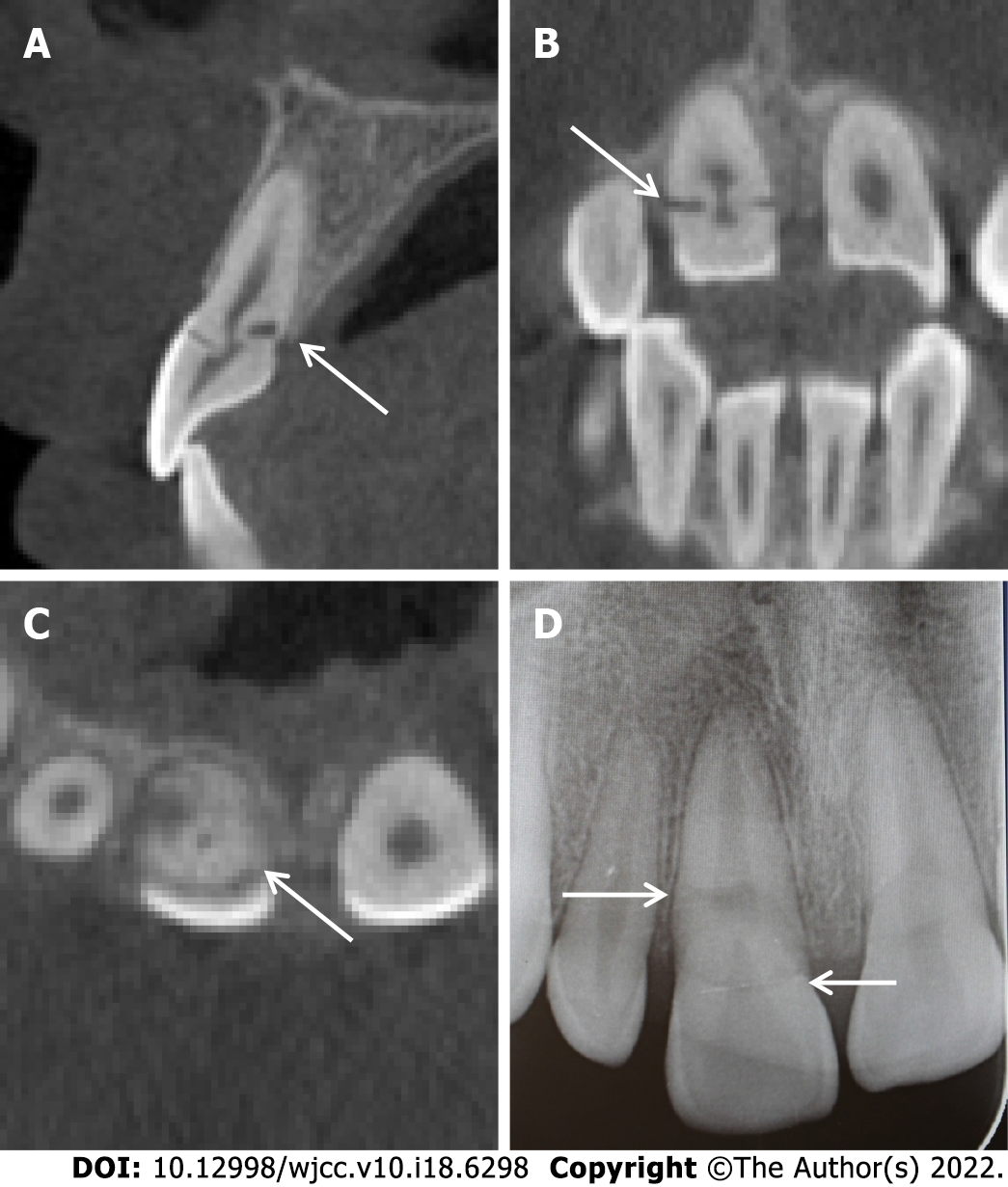
Figure 2 Cone beam computed tomography and periapical radiograph images showing an oblique crown-root fracture from the labial surface of the tooth 11 to the palatal alveolar ridge, which had signs of healing.
A: Sagittal; B: Coronal; C: Cross-sectional; D: Periapical.
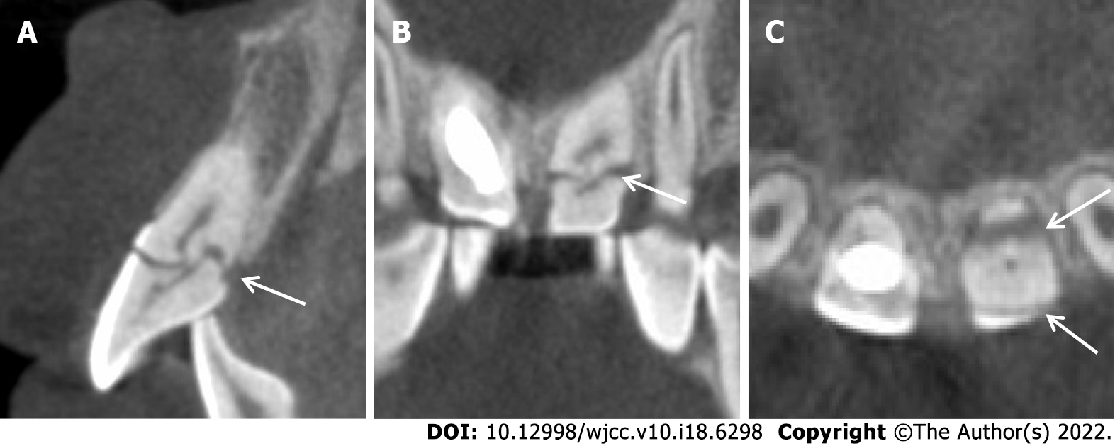
Figure 3 Cone beam computed tomography images showing an oblique crown-root fracture from the labial surface of the tooth 21 with hard tissue deposition at the pulpal side across the fracture line.
A: Sagittal; B: Coronal; C: Cross-sectional.
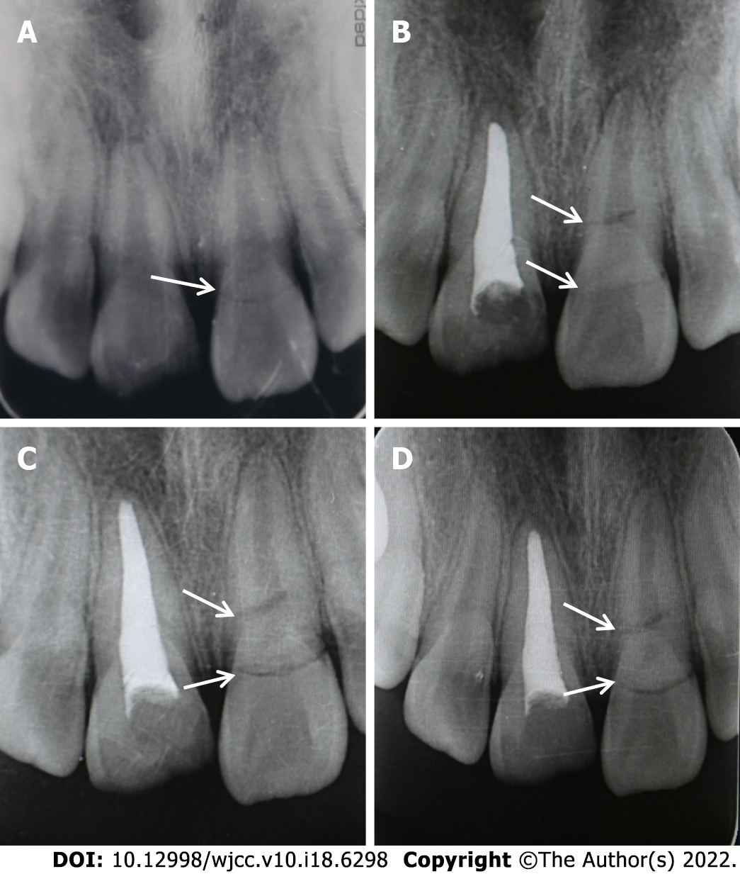
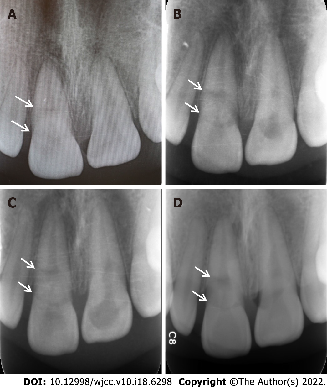
Figure 4 Periapical radiographs.
A: Periapical radiograph of teeth 11 and 21 after the first injury 1 yr ago showed fracture on tooth 21; B-D: Periapical radiographs of teeth 11 and 21 after tooth 11 was treated with apexification. The fracture line on tooth 21 became more evident over the time (B: 3 mo after initial injury; C: 6 mo after initial injury; D: 9 mo after initial injury).
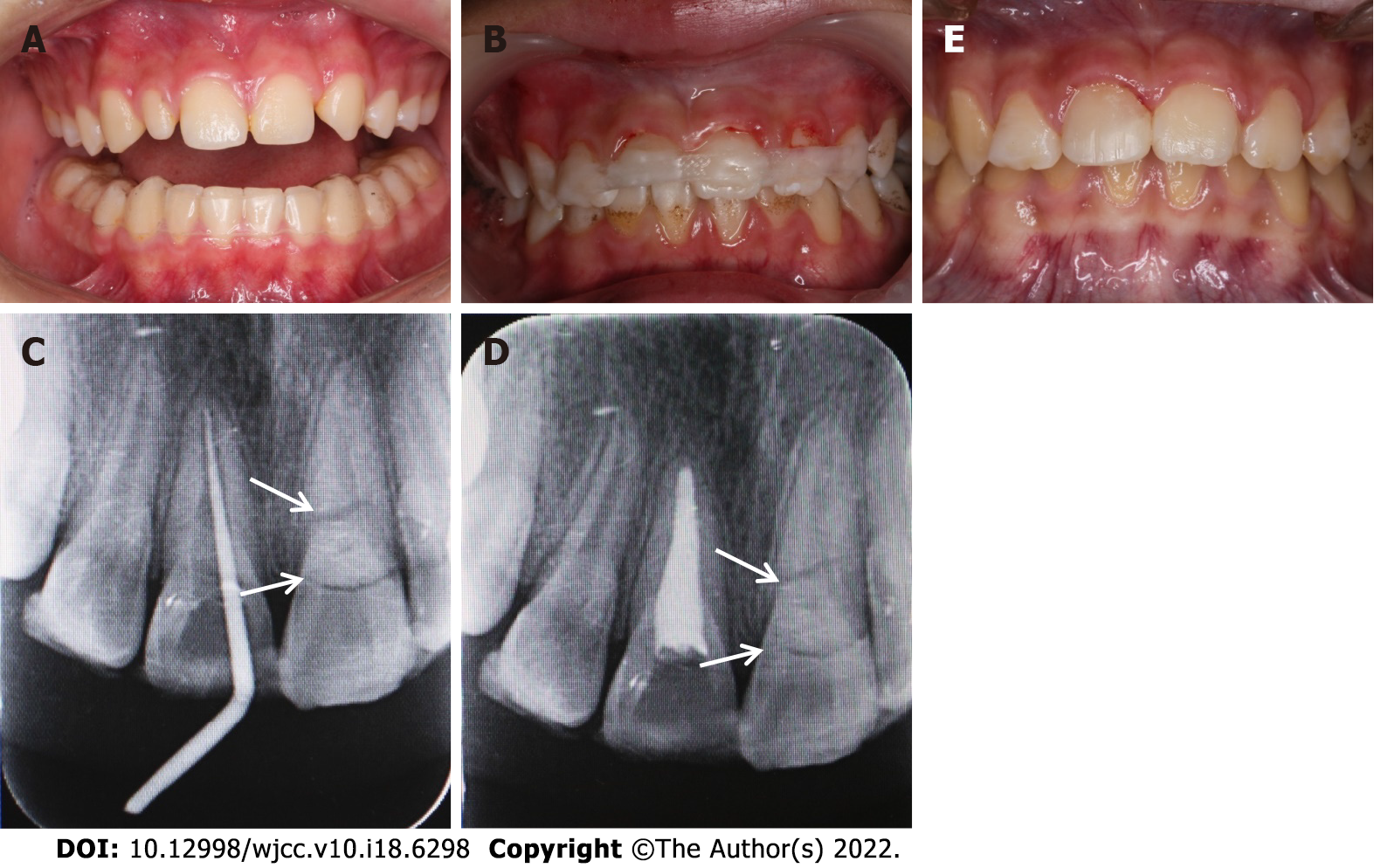
Figure 5 Treatment photographs.
A: Intraoral photograph after wearing a mandible occlusal pad; B: Tooth 21 was stabilized with a fiber splint; C and D: Completion of root canal therapy for tooth 11; E: Tooth 11 was restored with resin composite.
Figure 6 Periapical radiographs.
A-D: Periapical radiographs of tooth 11 taken 1 (A), 2 (B), 3 (C), and 4 yr later (D).
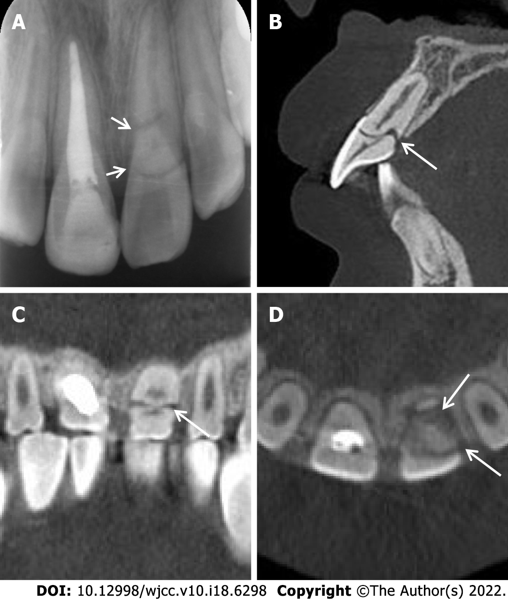
Figure 7 Periapical radiograph and cone beam computed tomography images.
A: Periapical radiograph of teeth 11 and 21 taken 9 mo after the restoration of tooth 11; B-D: Sagittal (B), coronal (C), and cross-sectional (D) cone beam computed tomography images taken 4 yr later showing sign of repair.
- Citation: Zhou ZL, Gao L, Sun SK, Li HS, Zhang CD, Kou WW, Xu Z, Wu LA. Spontaneous healing of complicated crown-root fractures in children: Two case reports. World J Clin Cases 2022; 10(18): 6298-6306
- URL: https://www.wjgnet.com/2307-8960/full/v10/i18/6298.htm
- DOI: https://dx.doi.org/10.12998/wjcc.v10.i18.6298


