Copyright
©The Author(s) 2022.
World J Clin Cases. Jun 26, 2022; 10(18): 6247-6253
Published online Jun 26, 2022. doi: 10.12998/wjcc.v10.i18.6247
Published online Jun 26, 2022. doi: 10.12998/wjcc.v10.i18.6247
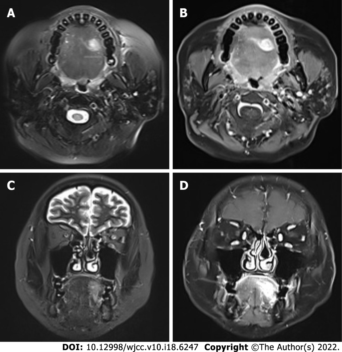
Figure 1 Magnetic resonance imaging indicated an abnormal signal intensity on the left side of the tongue.
A-D: T2-weighted magnetic resonance imaging (MRI) revealed a hyperintense shadow and contrast-enhanced T1-weighted MRI obvious enhancement of the mass in the transverse (A and B) and coronal planes (C and D). Scale bar: 2 cm.
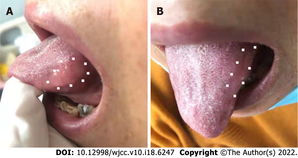
Figure 2 Irregular ill-defined nodule on the left side.
A-B: The tongue was of normal color on visual clinical examination in sagittal section (A) and transverse section (B). The dots indicate the margins of the nodule.
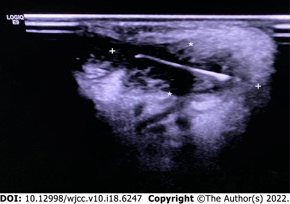
Figure 3 Ultrasound examination of the tongue.
Ultrasonography revealed an object of hyperechoic linear density, suggestive of an embedded foreign body (stars).
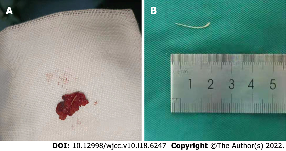
Figure 4 The foreign body.
A: Total enucleation without removal of the fish bone; B: The fish bone after removal.
- Citation: Jiang ZH, Xv R, Xia L. Foreign body granuloma in the tongue differentiated from tongue cancer: A case report. World J Clin Cases 2022; 10(18): 6247-6253
- URL: https://www.wjgnet.com/2307-8960/full/v10/i18/6247.htm
- DOI: https://dx.doi.org/10.12998/wjcc.v10.i18.6247


