Copyright
©The Author(s) 2022.
World J Clin Cases. Jun 26, 2022; 10(18): 6205-6210
Published online Jun 26, 2022. doi: 10.12998/wjcc.v10.i18.6205
Published online Jun 26, 2022. doi: 10.12998/wjcc.v10.i18.6205
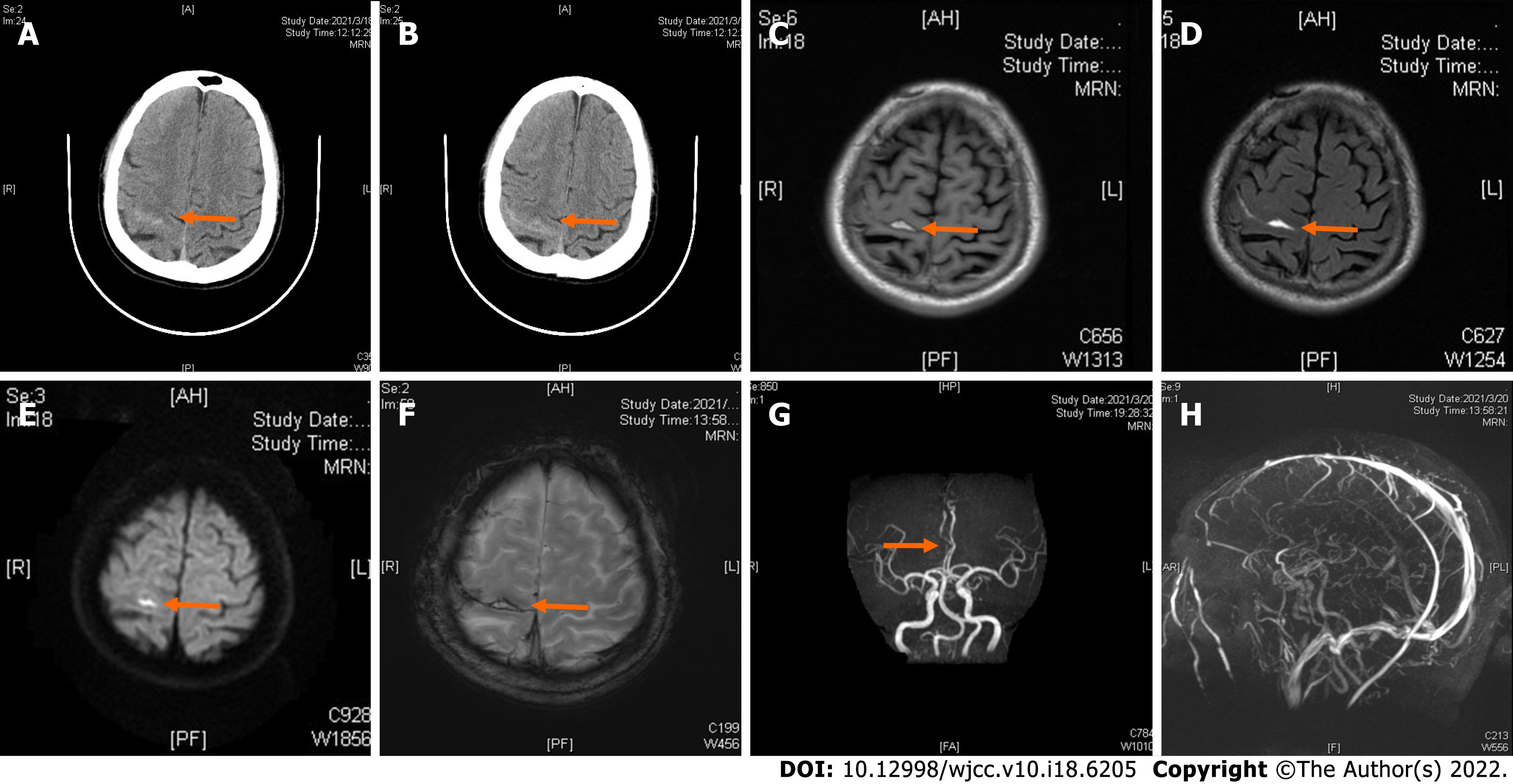
Figure 1 Computed tomography imaging.
A and B: Axial computed tomography images showing a high-density image of right frontal-parietal sulcus; C: Magnetic resonance imaging showing slightly elevated T1-flair; D: Elevated T2-flair; E: Diffusion-weighted imaging revealed high signal intensity; F: Susceptibility weighted imaging showing slightly increased signal intensity in the right frontal; G: Machine records activity showing short local stenosis of the right anterior cerebral artery of A3 segment; H: Magnetic resonance venography revealed a thinner contrast in the left transverse sinus and left sigmoid sinus.
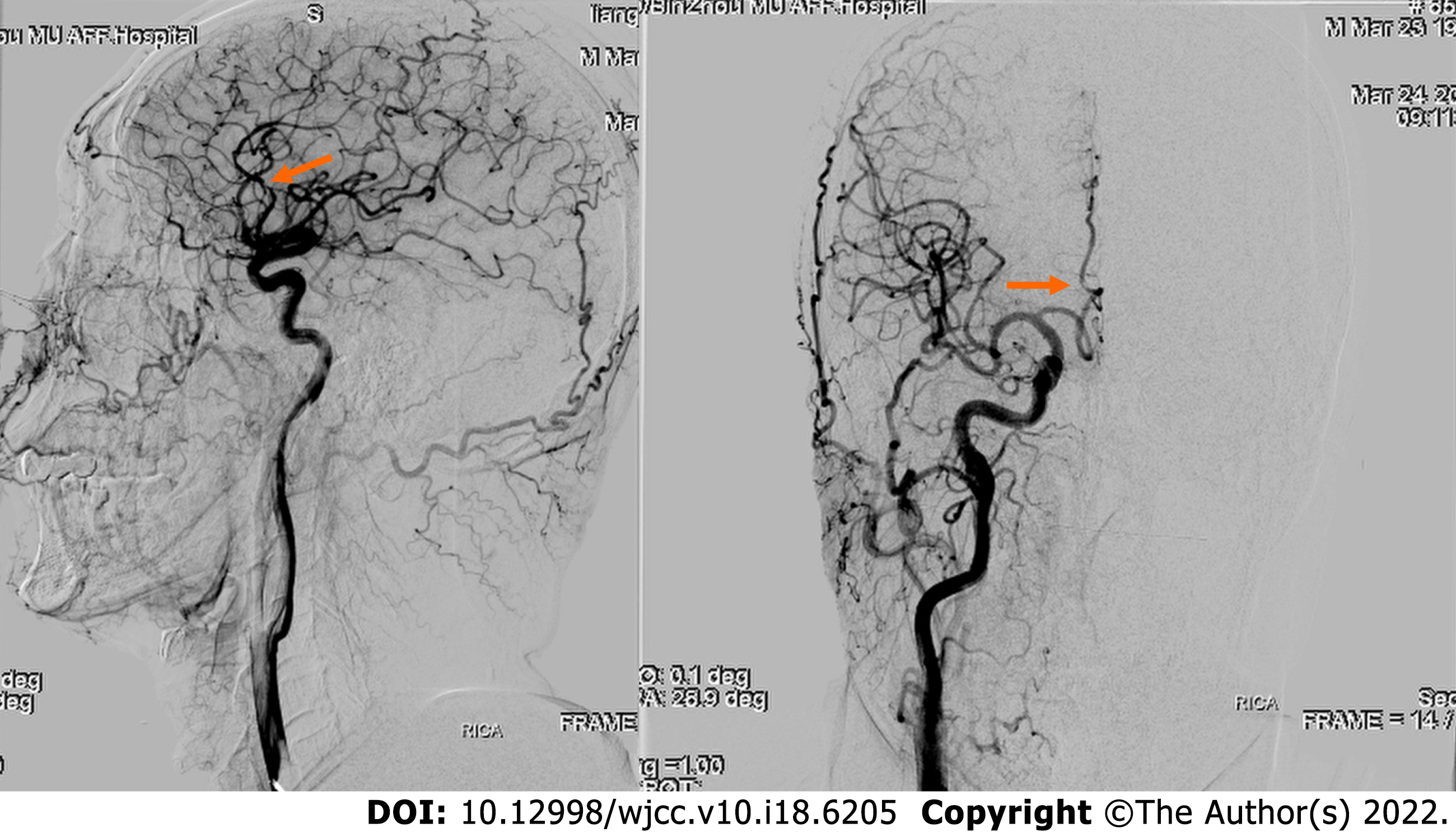
Figure 2 Digital subtraction angiography showing severe stenosis in the right anterior cerebral artery A2-A3 junction.
- Citation: Chen HL, Li B, Chen C, Fan XX, Ma WB. Nontraumatic convexal subarachnoid hemorrhage: A case report. World J Clin Cases 2022; 10(18): 6205-6210
- URL: https://www.wjgnet.com/2307-8960/full/v10/i18/6205.htm
- DOI: https://dx.doi.org/10.12998/wjcc.v10.i18.6205


