Copyright
©The Author(s) 2022.
World J Clin Cases. Jun 26, 2022; 10(18): 6141-6147
Published online Jun 26, 2022. doi: 10.12998/wjcc.v10.i18.6141
Published online Jun 26, 2022. doi: 10.12998/wjcc.v10.i18.6141
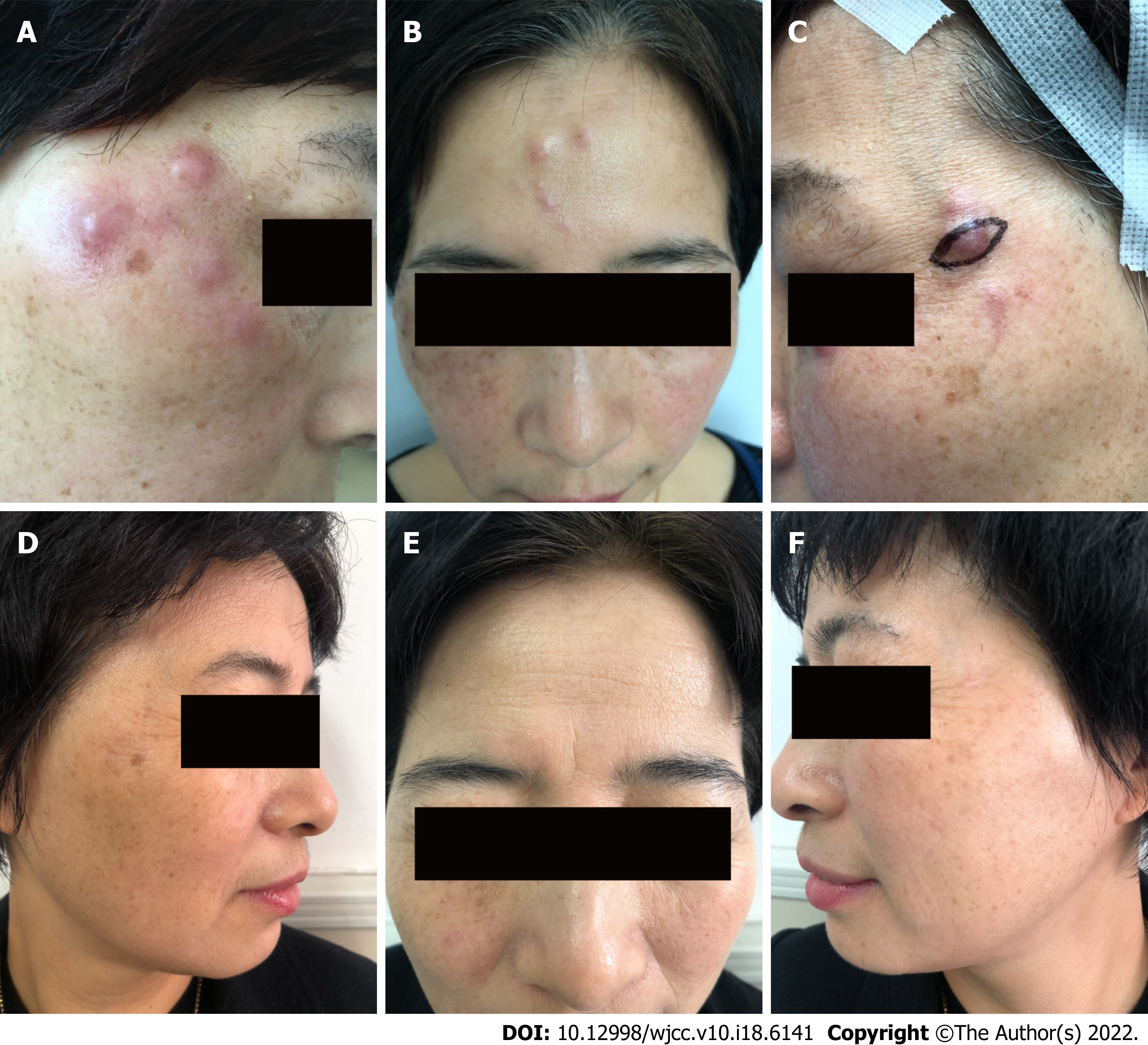
Figure 1 Patient’s photographs before and after treatment.
A-C: Multiple red papules, nodules, and abscesses were seen on the forehead and both temporal sites with a diameter of 1 cm-3 cm; D-F: The lesions were cured after the treatment for 7 mo.
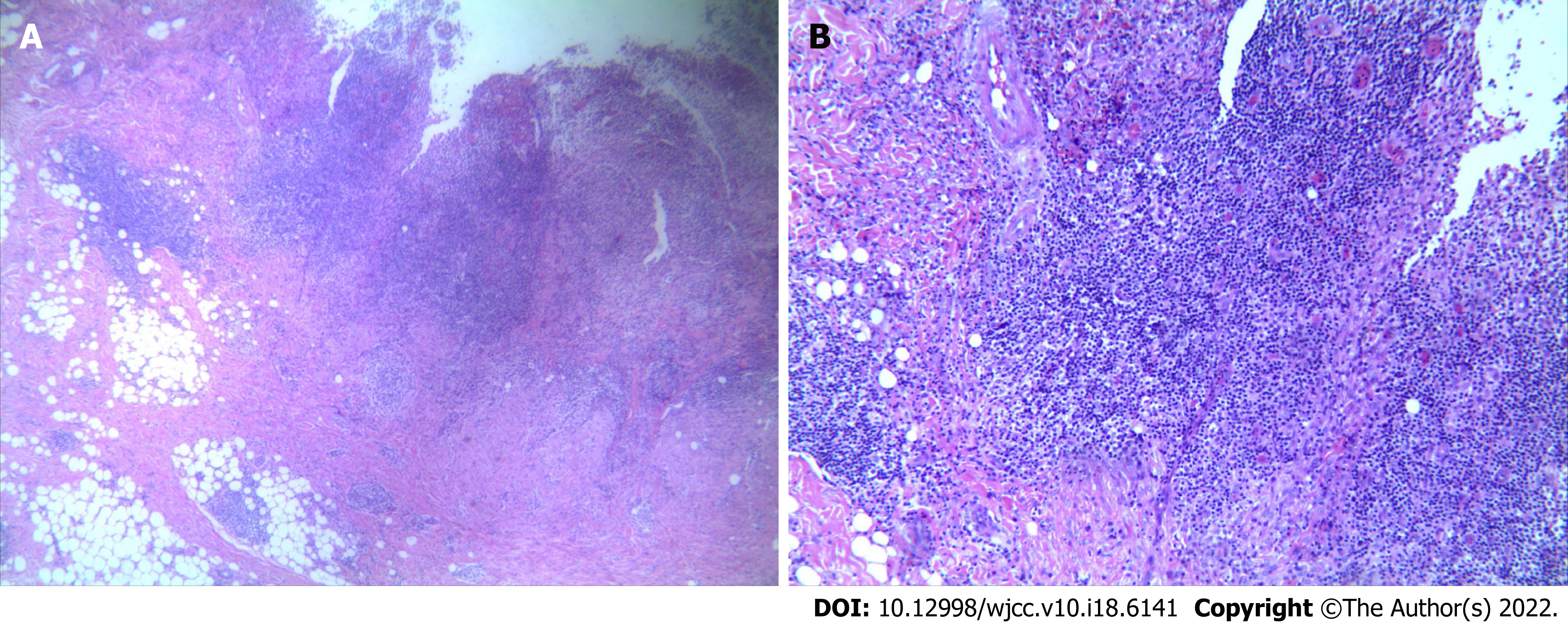
Figure 2 Histopathology.
The histopathology indicated a large number of mixed inflammatory cell infiltrates in the deep dermis, including neutrophils, histiocytes, and lymphocytes (A: HE × 40, B: HE × 100).
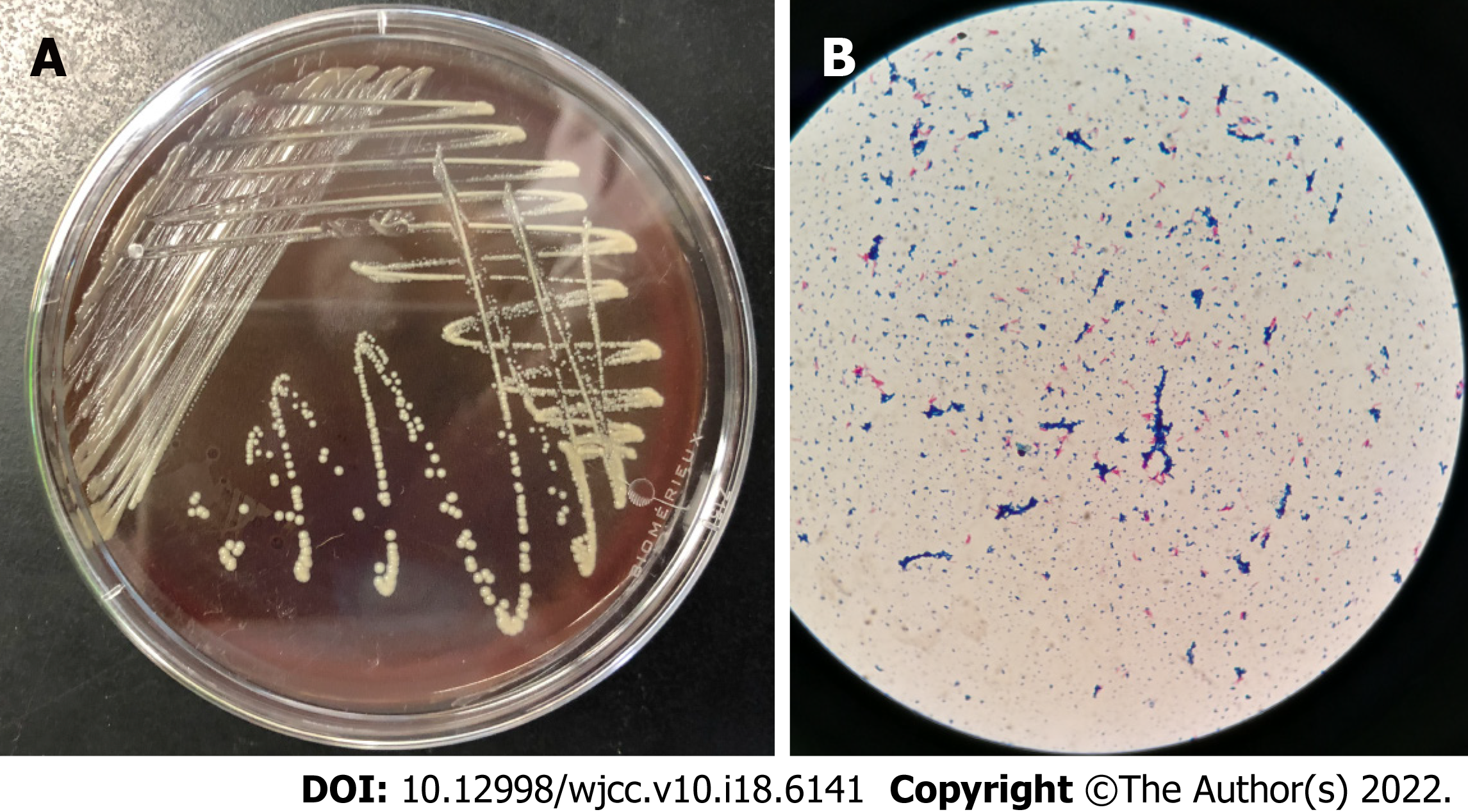
Figure 3 Microbiological evidence.
A: Cultures on Mycobacterium Roche's Medium yielded cream-colored, yeast-like colonies within 5 d at 35 °C; B: Scattered pink rod-shaped bacteria after acid-fast staining (× 100).
- Citation: Deng L, Luo YZ, Liu F, Yu XH. Subcutaneous infection caused by Mycobacterium abscessus following cosmetic injections of botulinum toxin: A case report. World J Clin Cases 2022; 10(18): 6141-6147
- URL: https://www.wjgnet.com/2307-8960/full/v10/i18/6141.htm
- DOI: https://dx.doi.org/10.12998/wjcc.v10.i18.6141


