Copyright
©The Author(s) 2022.
World J Clin Cases. Jun 26, 2022; 10(18): 6105-6109
Published online Jun 26, 2022. doi: 10.12998/wjcc.v10.i18.6105
Published online Jun 26, 2022. doi: 10.12998/wjcc.v10.i18.6105
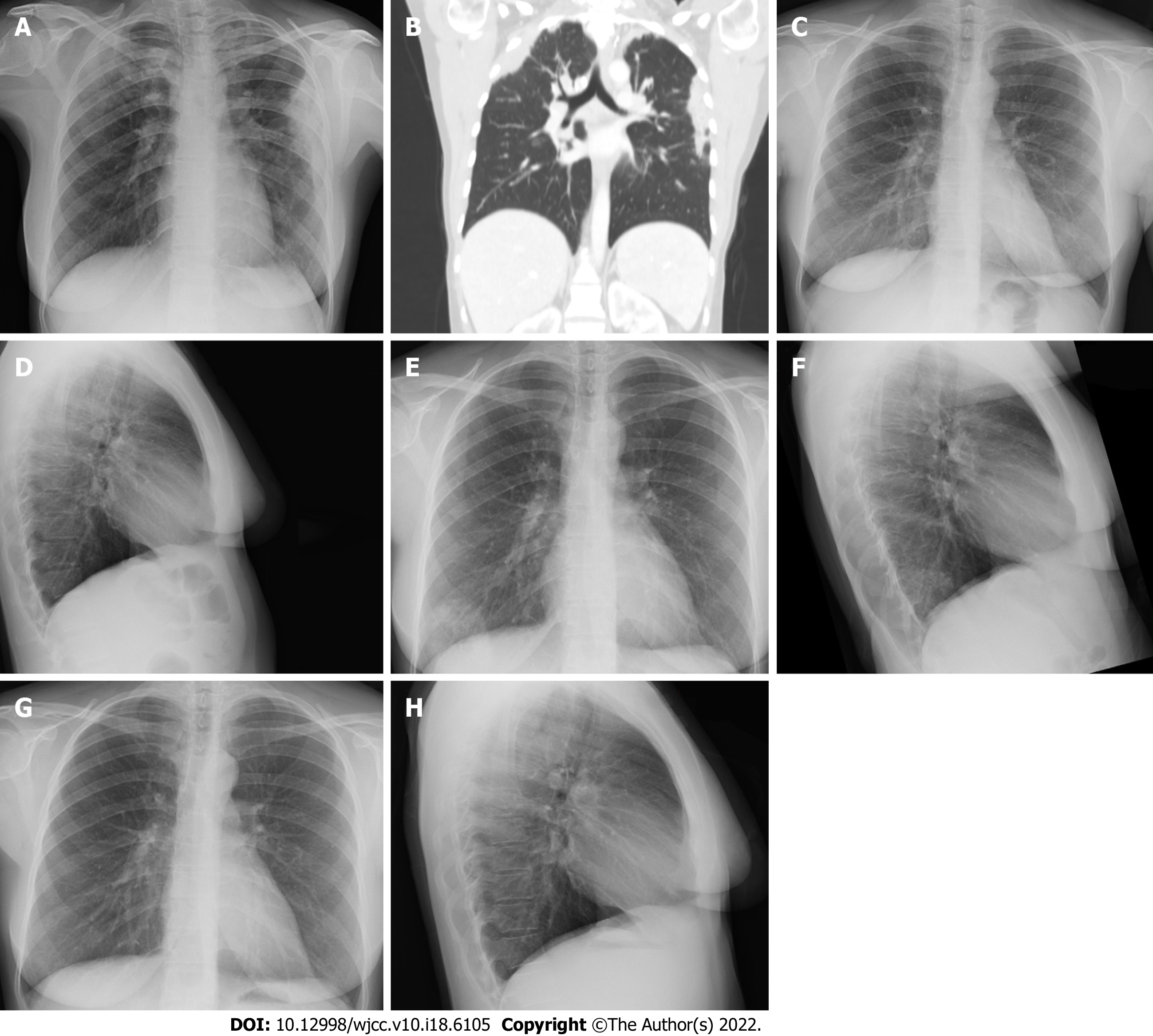
Figure 1 Imaging examinations of the present patient.
A: Chest X-ray (frontal view) shows infiltrates in the right upper and left lateral lung fields (two weeks prior to the patient’s presentation in our hospital); B: CT of the chest (axial plain) reveals a mediastinal lymphadenopathy, and pulmonary consolidations in the right upper and left lower lobes (at first presentation); C: Chest X-ray (frontal view) and D: (lateral view) show disappearance of the pulmonary infiltrates (6 wk after the presentation); E: Chest X-ray (frontal view) and F: (lateral view) reveal a new infiltrate in the right lower lobe (4.5 mo after the presentation); G: Chest X-ray (frontal view) and H: (lateral view) reveal absorption of the infiltrate 5 wk after beginning of benralizumab treatment.
- Citation: Izhakian S, Pertzov B, Rosengarten D, Kramer MR. Successful treatment of acute relapse of chronic eosinophilic pneumonia with benralizumab and without corticosteroids: A case report . World J Clin Cases 2022; 10(18): 6105-6109
- URL: https://www.wjgnet.com/2307-8960/full/v10/i18/6105.htm
- DOI: https://dx.doi.org/10.12998/wjcc.v10.i18.6105


