Copyright
©The Author(s) 2022.
World J Clin Cases. Jun 26, 2022; 10(18): 6039-6049
Published online Jun 26, 2022. doi: 10.12998/wjcc.v10.i18.6039
Published online Jun 26, 2022. doi: 10.12998/wjcc.v10.i18.6039
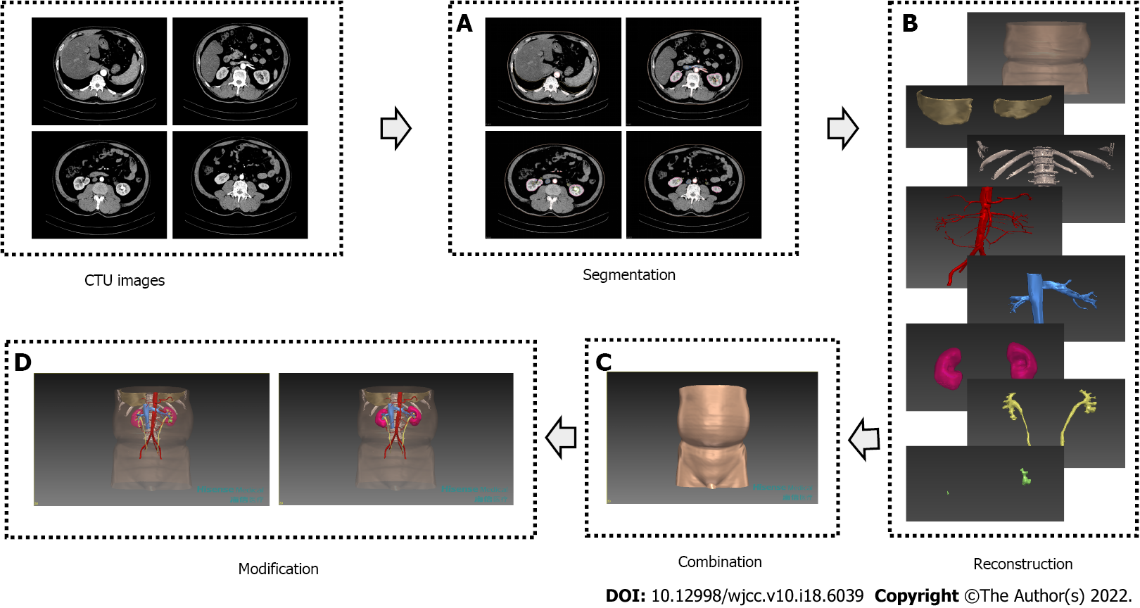
Figure 1 The procedures of three-dimensional model construction by the computer-assisted surgery system.
A: Different structures were segmented from different phases of CTU; B: Segmented skin, the bottom of the thoracic cavity, spine, ribs, renal arterial system, renal venous system, renal parenchyma, renal collecting system, ureters and stones were reconstructed respectively; C: In combination, reconstructed structures were registered and integrated into a fusion model; D: In modification, diaphaneity adjustment of skin, renal parenchyma and renal collecting system made it possible to observe stones.
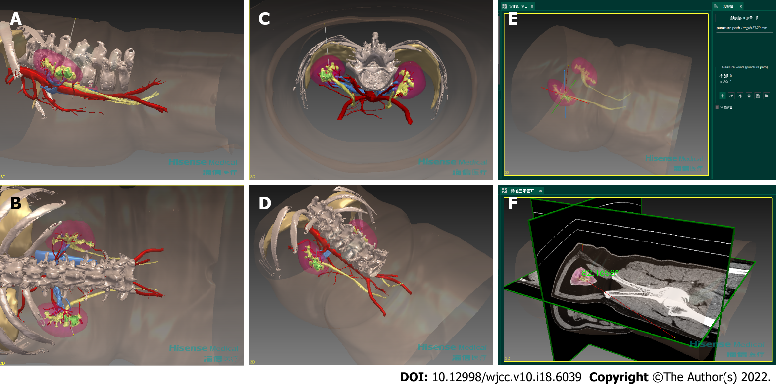
Figure 2 Preoperative planning for percutaneous tracts with the three-dimensional model.
Ideal puncture path in side view (A), back view (B), axial view (C) and oblique view (D) were simulated. Lengths and angles could be measured by the CAS system, taking the depth of ideal puncture path (E) and the angle between ideal puncture path and the coronal plane (F) for example.
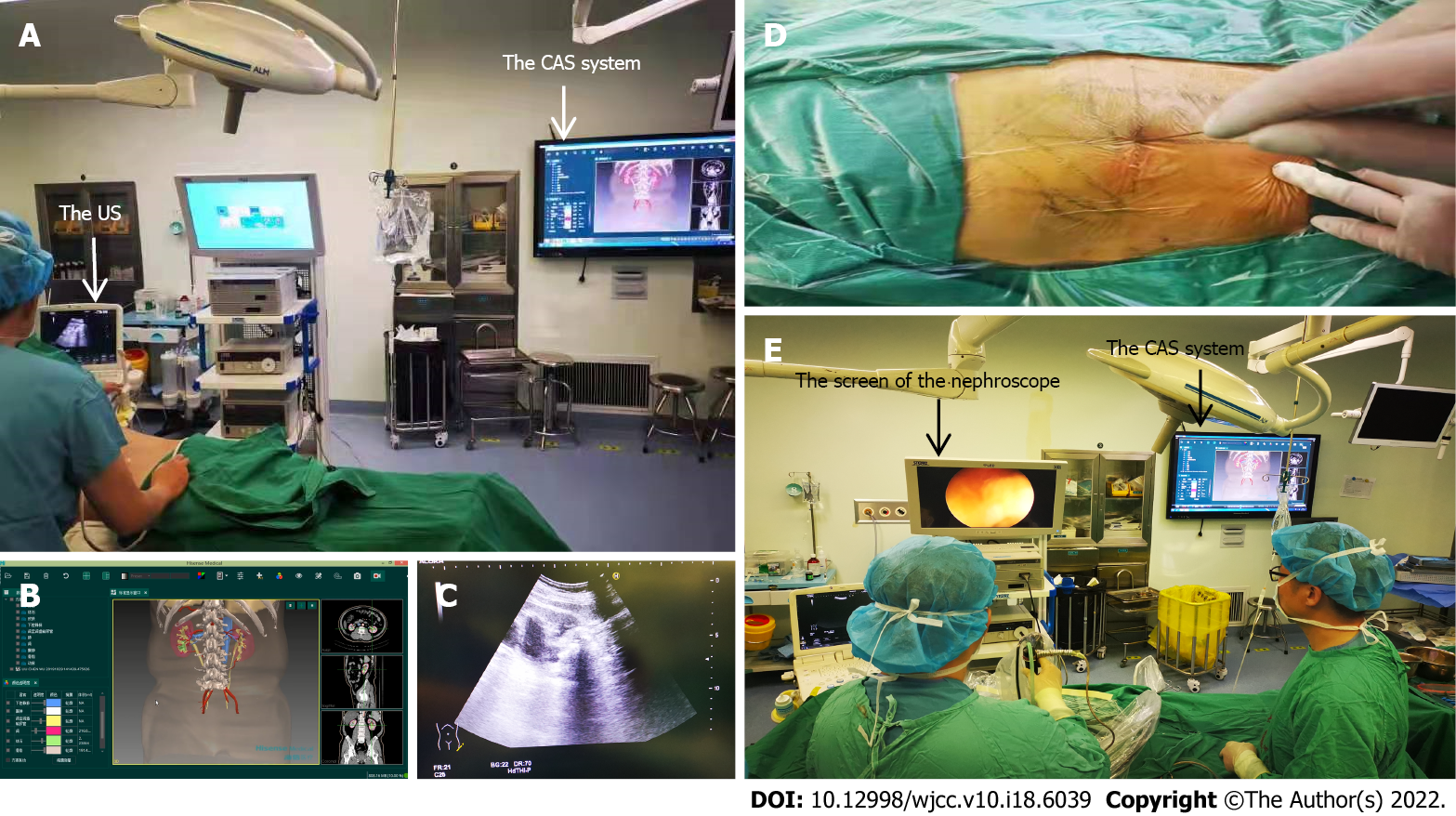
Figure 3 Intraoperative navigation for percutaneous nephrolithotomy with the computer-assisted surgery system.
A: Making percutaneous tracts with the help of the CAS system and the US; B: The CAS system showed the 3D model and the plan obtained preoperatively; C: The US showed puncture paths and surrounding structures in real time; D: Successful puncture was guided by parameters from preoperative planning; E: Removing stones with the reference from the CAS system.
- Citation: Qin F, Sun YF, Wang XN, Li B, Zhang ZL, Zhang MX, Xie F, Liu SH, Wang ZJ, Cao YC, Jiao W. Application of a novel computer-assisted surgery system in percutaneous nephrolithotomy: A controlled study. World J Clin Cases 2022; 10(18): 6039-6049
- URL: https://www.wjgnet.com/2307-8960/full/v10/i18/6039.htm
- DOI: https://dx.doi.org/10.12998/wjcc.v10.i18.6039


