Copyright
©The Author(s) 2022.
World J Clin Cases. Jun 26, 2022; 10(18): 6032-6038
Published online Jun 26, 2022. doi: 10.12998/wjcc.v10.i18.6032
Published online Jun 26, 2022. doi: 10.12998/wjcc.v10.i18.6032
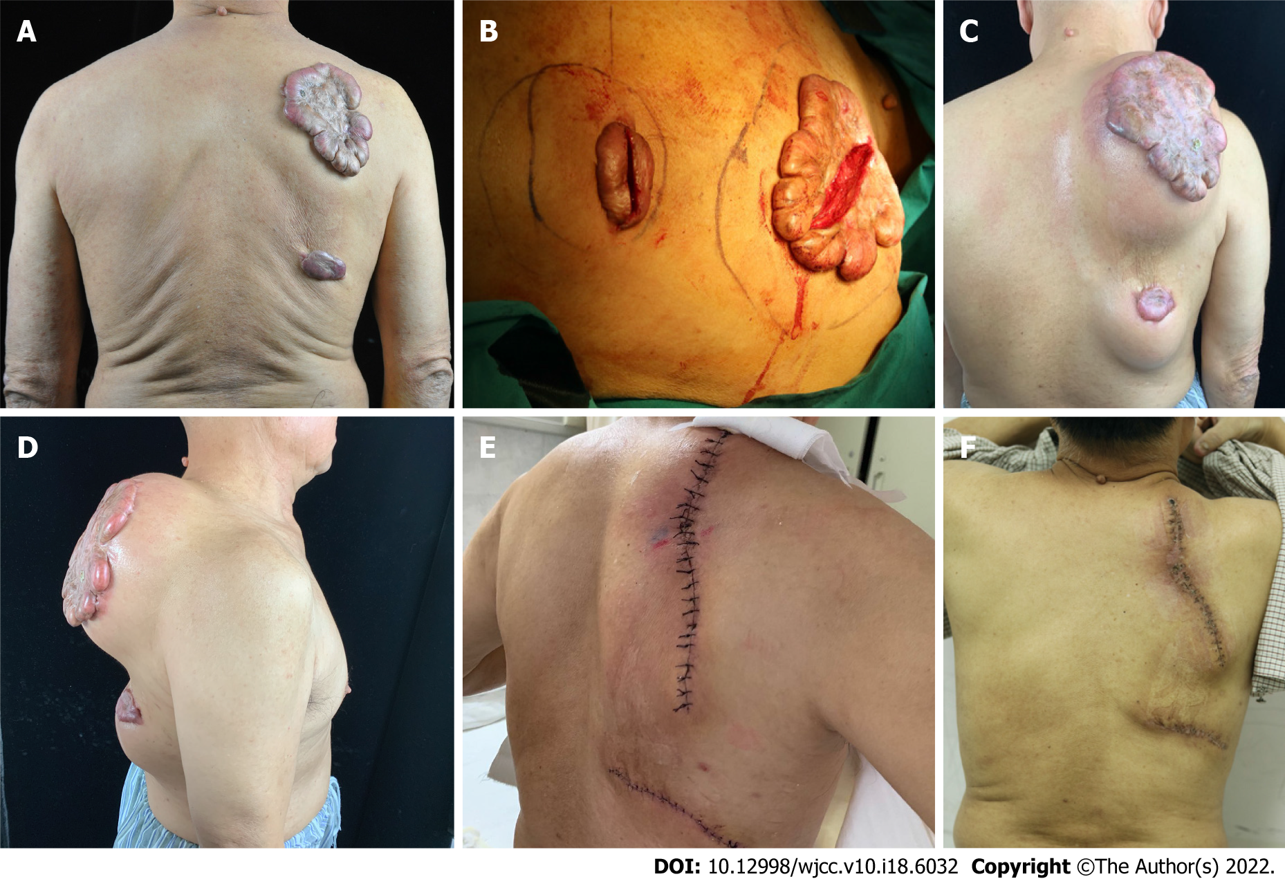
Figure 1 A 69-year-old patient with keloids.
A: Two prominent skin, and the edge beyond the scar base; B: In the first stage of operation, expander implantation was performed through an intrascar incision; C, D: The methylene blue line was the designed range of expander implantation; after expander implantation, normal saline was rapidly injected to dilate it; expander water injection; E: After injection of saline, the expander was removed and the expanded flap was sutured. Continuous superficial radiotherapy after the operation, for 4 d; F: Two weeks after the operation, immediate suture removal was done.
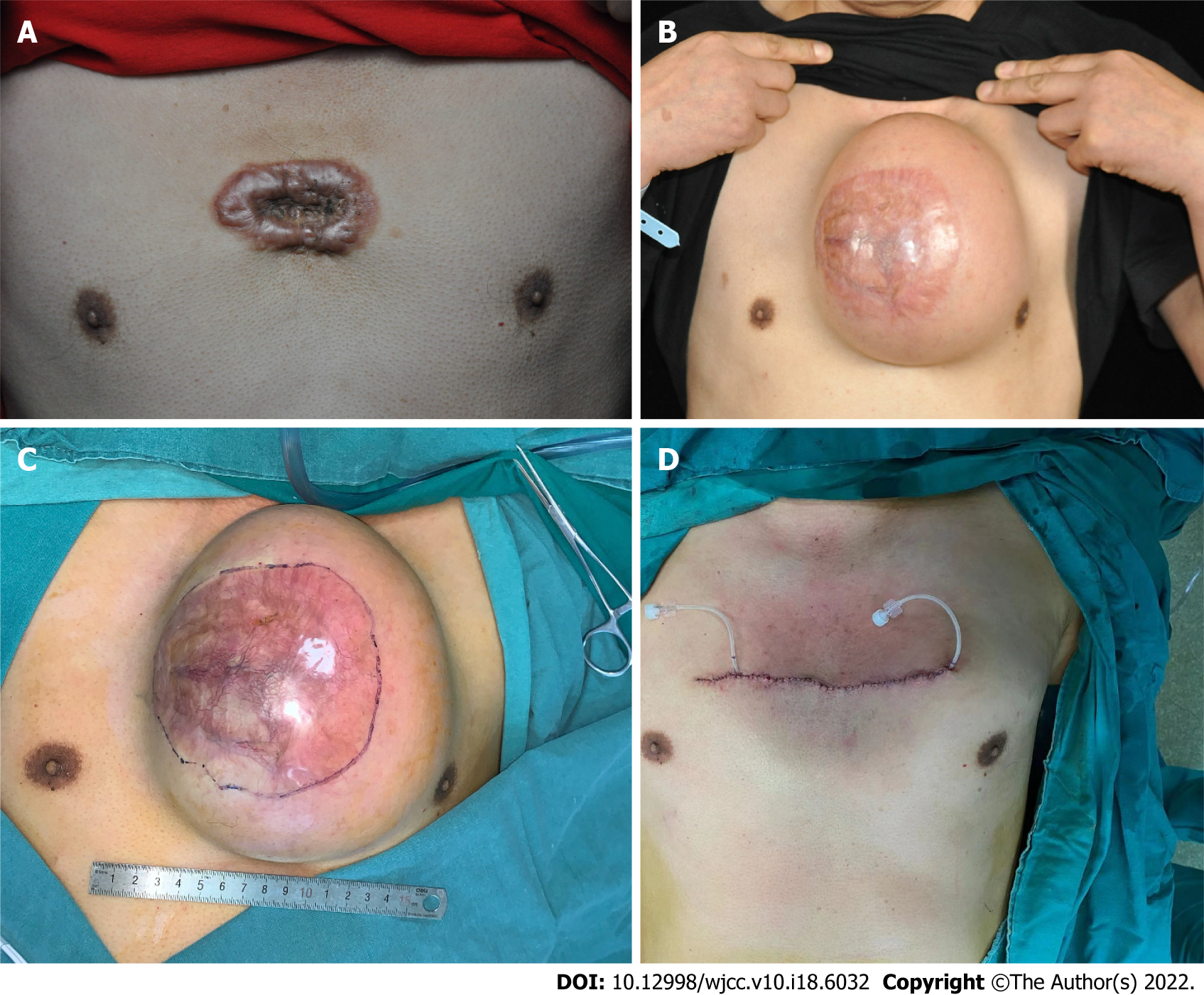
Figure 2 A patient with an anterior chest keloid.
A: The patient with an anterior chest keloid, prominent skin lesions, and edge-invasive growth appearance; B: Scar incision after expander implantation and normal saline expansion; C: During expander removal, methylene blue was designed to remove keloid size; D: Immediately after scar excision and expander removal.
- Citation: Wu M, Gu JY, Duan R, Wei BX, Xie F. Scar-centered dilation in the treatment of large keloids. World J Clin Cases 2022; 10(18): 6032-6038
- URL: https://www.wjgnet.com/2307-8960/full/v10/i18/6032.htm
- DOI: https://dx.doi.org/10.12998/wjcc.v10.i18.6032


