Copyright
©The Author(s) 2022.
World J Clin Cases. Jun 16, 2022; 10(17): 5825-5832
Published online Jun 16, 2022. doi: 10.12998/wjcc.v10.i17.5825
Published online Jun 16, 2022. doi: 10.12998/wjcc.v10.i17.5825
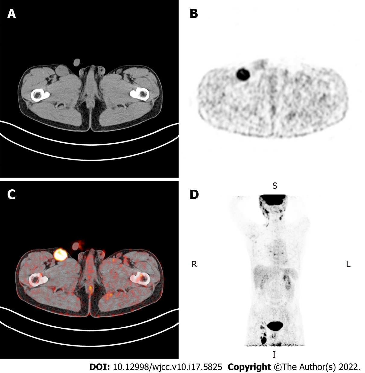
Figure 1 Positron emission tomography-computed tomography examination.
A: Computed tomography (CT) examination detected enlarged lymph node in the right inguinal region; B: Positron emission tomography (PET) images showed that metabolism was obviously increased in the enlarged lymph node; C: PET-CT fusion image; D: The image of PET in the coronal plane indicated abnormal fluorodeoxyglucose accumulation in the whole body.
Figure 2 Orbital computed tomography.
A: The orbital computed tomography (CT) image in June 2019 showed slightly swelling surrounded the eyes; B: Preoperative orbital CT in April 2020 indicated that left eyeball and extra-ocular muscle were compressed; C: Postoperative orbital CT 3 mo after microwave ablation.
Figure 3 Preoperative sonography of the eyelid mass.
A: Preoperative ultrasonography of the eyelid mass; B: Colour Doppler flow imaging showed the mass was rich in blood flow signals; C: Marked contrast enhancement of the mass was observed via contrast-enhanced ultrasound.
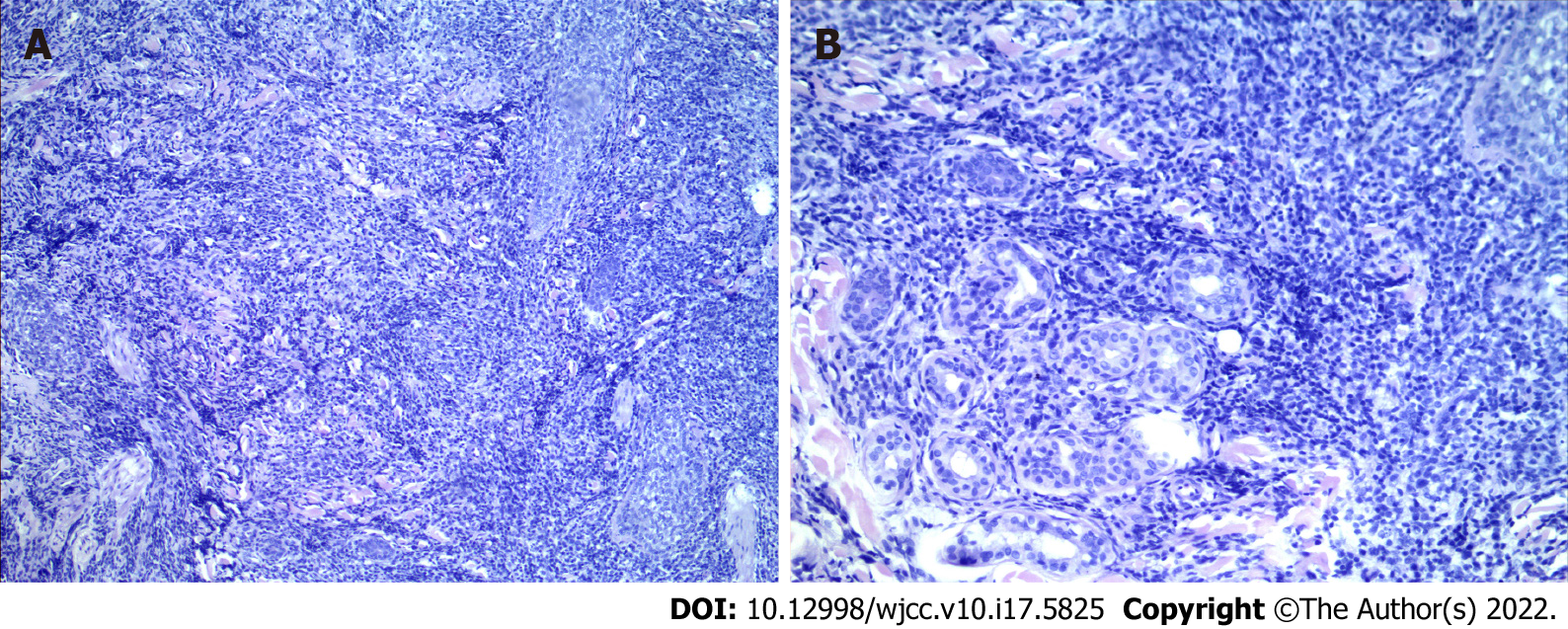
Figure 4 Pathological examination of the facial skin.
A: Magnification: 200 ×; B: Magnification: 400 ×.
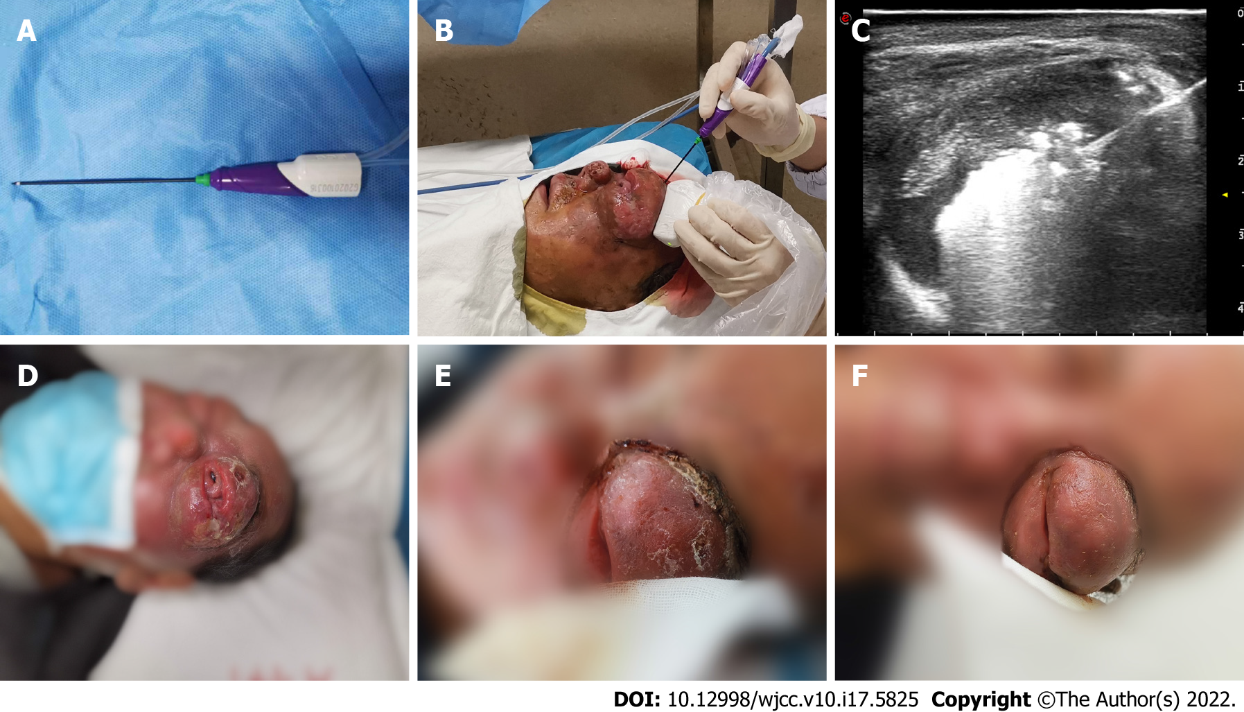
Figure 5 Procedure and follow-up of microwave ablation.
A: The disposable microwave therapeutic antenna; B: The microwave ablation was performed under ultrasound guidance; C: Ultrasound image showed microwave energy was being released; D: One day before microwave ablation; E: One week after microwave ablation; F: Two weeks after microwave ablation.
- Citation: Chen YW, Yang HZ, Zhao SS, Zhang Z, Chen ZM, Feng HH, An MH, Wang KK, Duan R, Chen BD. Ultrasound-guided microwave ablation as a palliative treatment for mycosis fungoides eyelid involvement: A case report. World J Clin Cases 2022; 10(17): 5825-5832
- URL: https://www.wjgnet.com/2307-8960/full/v10/i17/5825.htm
- DOI: https://dx.doi.org/10.12998/wjcc.v10.i17.5825


