Copyright
©The Author(s) 2022.
World J Clin Cases. Jun 16, 2022; 10(17): 5789-5797
Published online Jun 16, 2022. doi: 10.12998/wjcc.v10.i17.5789
Published online Jun 16, 2022. doi: 10.12998/wjcc.v10.i17.5789
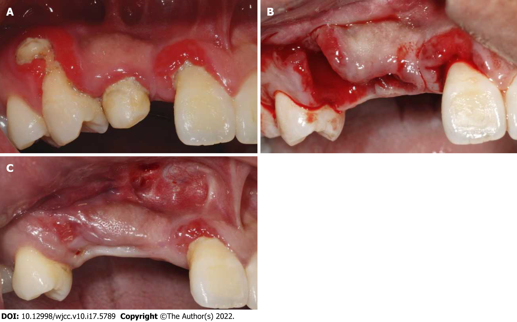
Figure 1 Oral examination of the patient at initial visit, after tooth extraction and before bone grafting.
A: Initial situation of the edentulous site in maxillary anterior; B: Extraction of teeth 53 and 14; C: Three weeks after teeth extraction and before bone augmentation surgery.
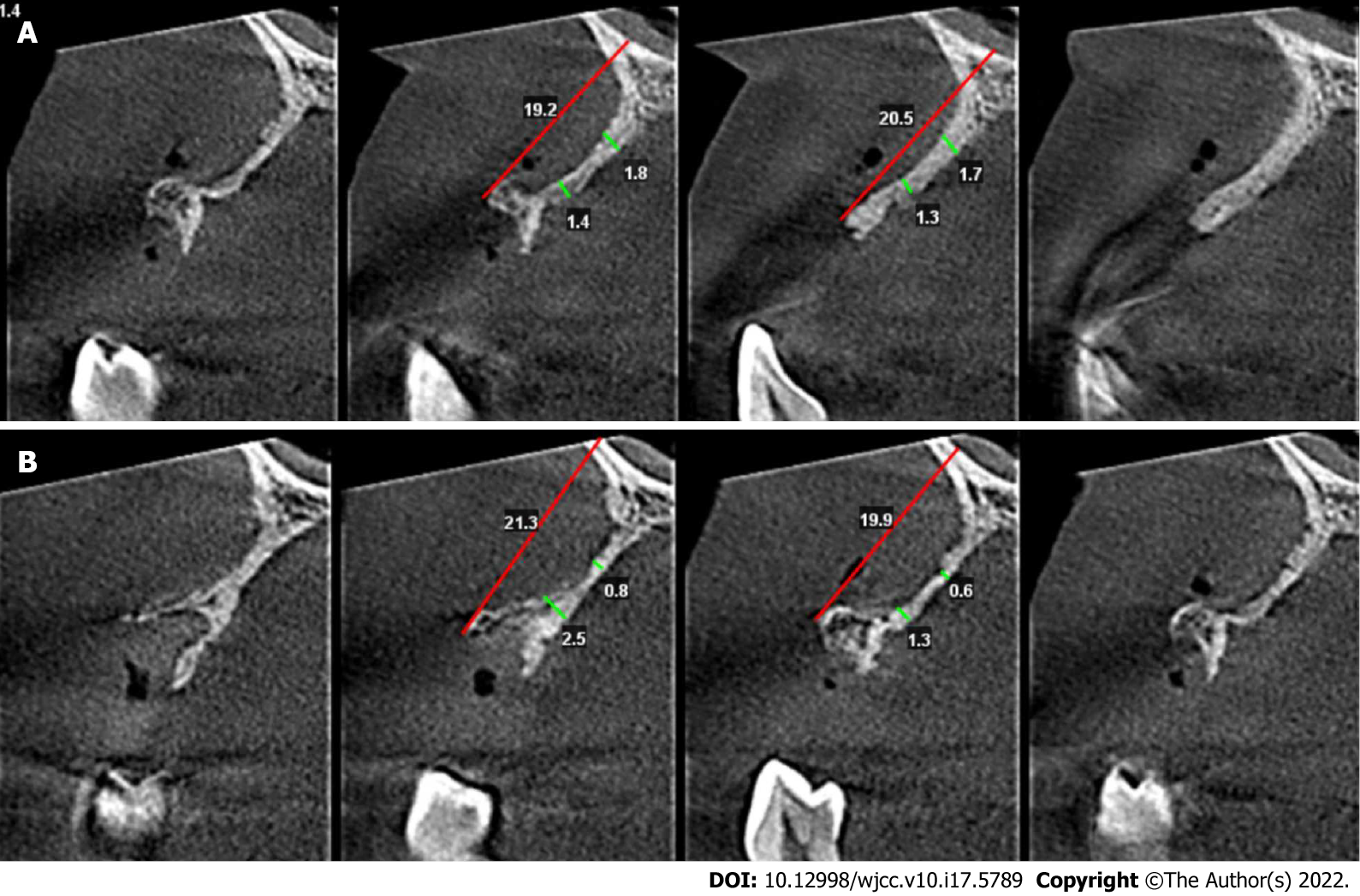
Figure 2 Bone volume at the defect area before bone grafting.
A: Cone beam computed tomography (CBCT) before bone augmentation surgery 5 and 10 mm below the alveolar crest at the site of missing tooth 12; B: CBCT before bone augmentation surgery 5 and 10 mm below the alveolar crest at the site of missing tooth 14.
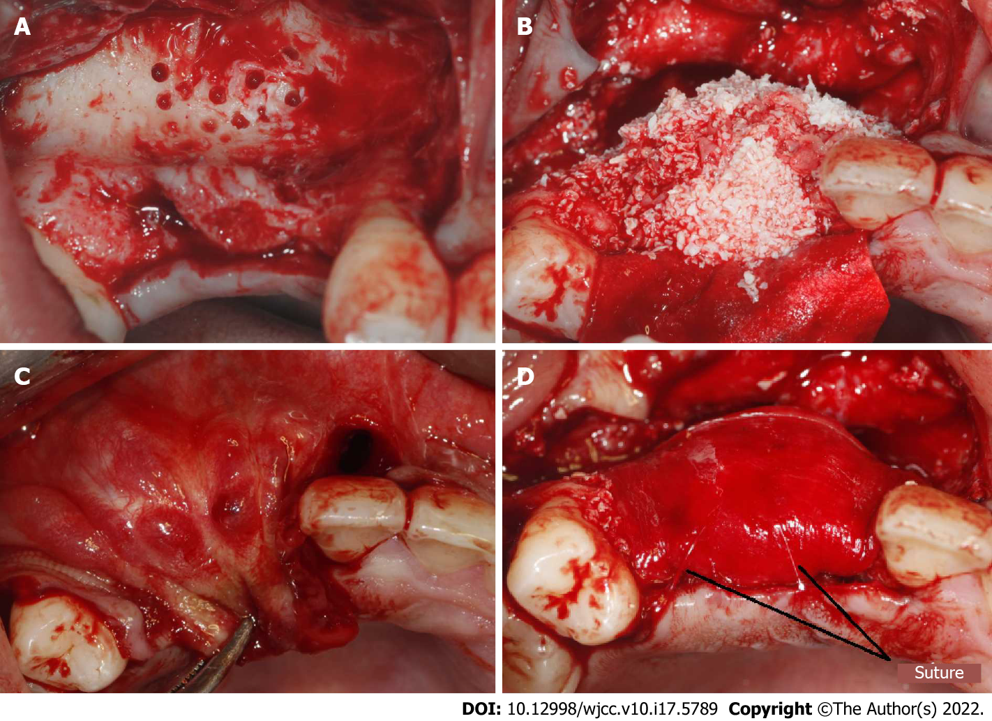
Figure 3 Procedure of bone grafting.
A: Decortication holes were prepared at the recipient area; B: Bone graft material was placed; C: Make sure the soft tissue could be primary closed without tension; D: The resorbable membranes and bone grafts were fixed using periosteal diagonal mattress sutures and four corner pins.
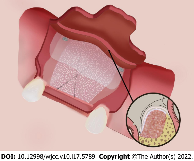
Figure 4 Fixation of the membrane using periosteal diagonal mattress suture and four corner pins.
Figure 5 Cone beam computed tomography superimposed images before bone grafting and after implantation.
A: At site of implant 12. The yellow line represented the alveolar ridge before bone grafting. The bone width was increased from 1.83 to 8.83 mm at a point 5 mm below the crest, and from 1.70 to 9.47 mm at a point 10 mm below the crest; B: At site of implant 14. The yellow line represented the alveolar ridge before bone grafting. The bone width was increased from 0.72 to 9.23 mm at a point 5 mm below the crest, and from 4.22 to 11.55 mm at a point 10 mm below the crest.
- Citation: Wang LH, Ruan Y, Zhao WY, Chen JP, Yang F. Modified membrane fixation technique in a severe continuous horizontal bone defect: A case report. World J Clin Cases 2022; 10(17): 5789-5797
- URL: https://www.wjgnet.com/2307-8960/full/v10/i17/5789.htm
- DOI: https://dx.doi.org/10.12998/wjcc.v10.i17.5789


