Copyright
©The Author(s) 2022.
World J Clin Cases. Jun 16, 2022; 10(17): 5783-5788
Published online Jun 16, 2022. doi: 10.12998/wjcc.v10.i17.5783
Published online Jun 16, 2022. doi: 10.12998/wjcc.v10.i17.5783
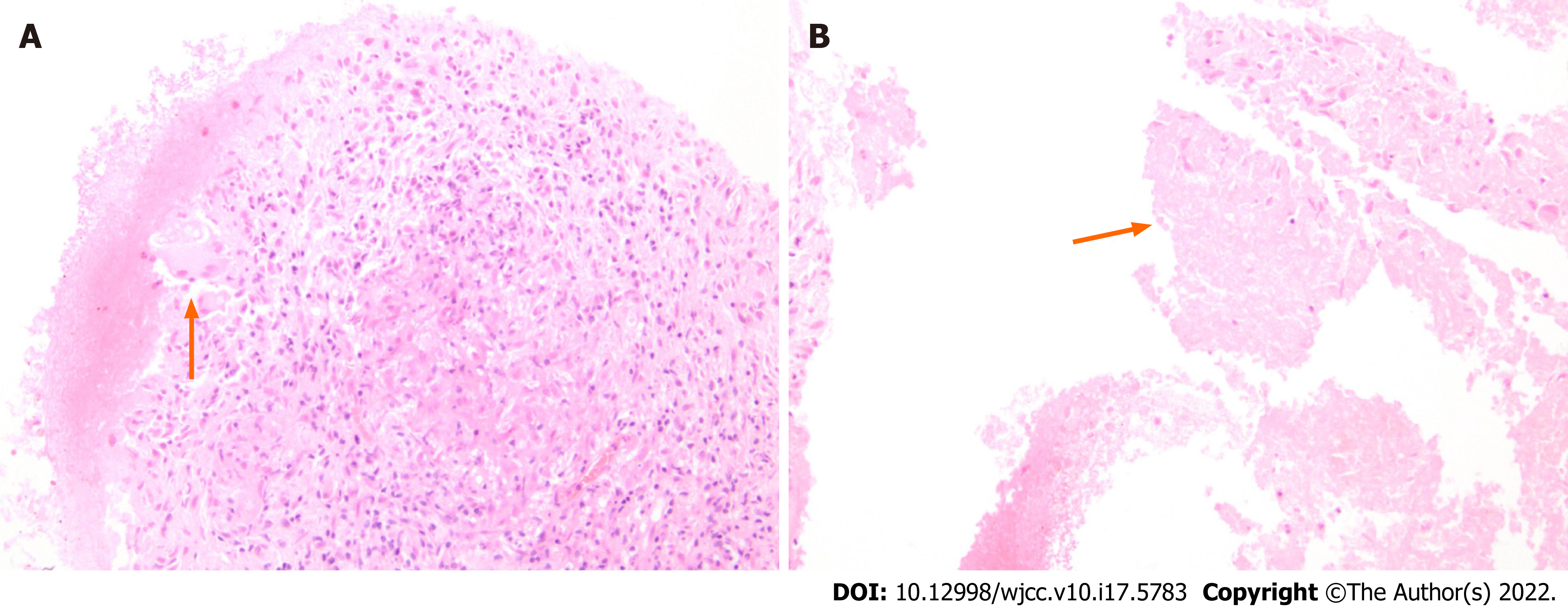
Figure 1 Microscopic examination of fine needle aspiration biopsy of the right adrenal gland showed Langerhans cells.
A: Epithelioid cells; B: Necrosis; which were suggestive of tuberculosis infection.
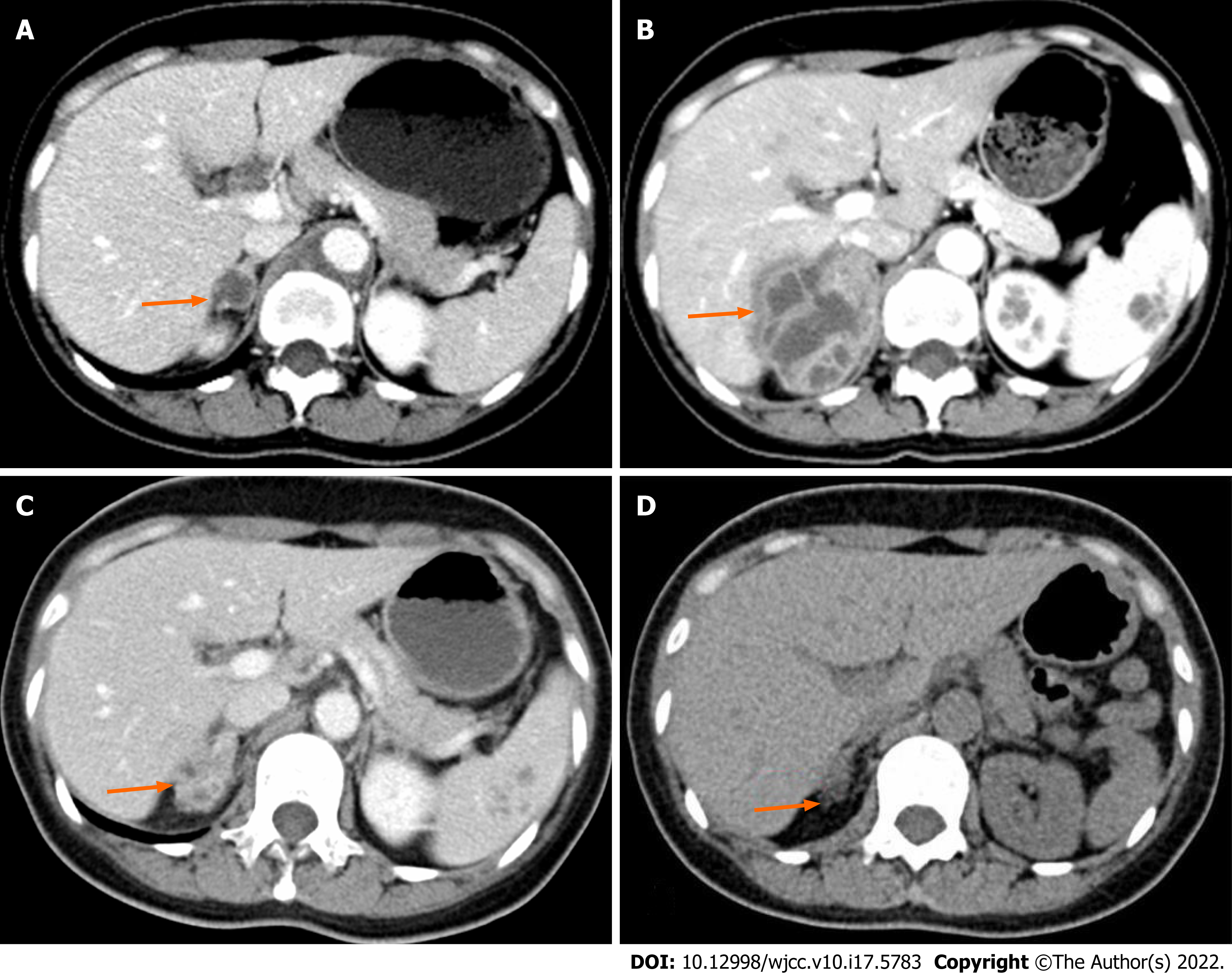
Figure 2 These were contrast-enhanced axial computed tomography images of the abdomen.
A: Showed an isointense mass of 1.5 cm in the right adrenal gland; B: Showed an irregular mass of 6.0 cm × 4.5 cm with uneven density (20-30 HU in the central and 80-90 HU in the peripheral) in the right adrenal gland, while the left one was normal; C: Showed that the mass became smaller (2.7 cm × 2.4 cm) after anti-TB therapy for a total of 15 mo; D: Showed a slight enlargement of the right adrenal gland after about three years’ anti-TB therapy.
- Citation: Liu H, Tang TJ, An ZM, Yu YR. Unilateral adrenal tuberculosis whose computed tomography imaging characteristics mimic a malignant tumor: A case report. World J Clin Cases 2022; 10(17): 5783-5788
- URL: https://www.wjgnet.com/2307-8960/full/v10/i17/5783.htm
- DOI: https://dx.doi.org/10.12998/wjcc.v10.i17.5783


