Copyright
©The Author(s) 2022.
World J Clin Cases. May 6, 2022; 10(13): 4280-4287
Published online May 6, 2022. doi: 10.12998/wjcc.v10.i13.4280
Published online May 6, 2022. doi: 10.12998/wjcc.v10.i13.4280
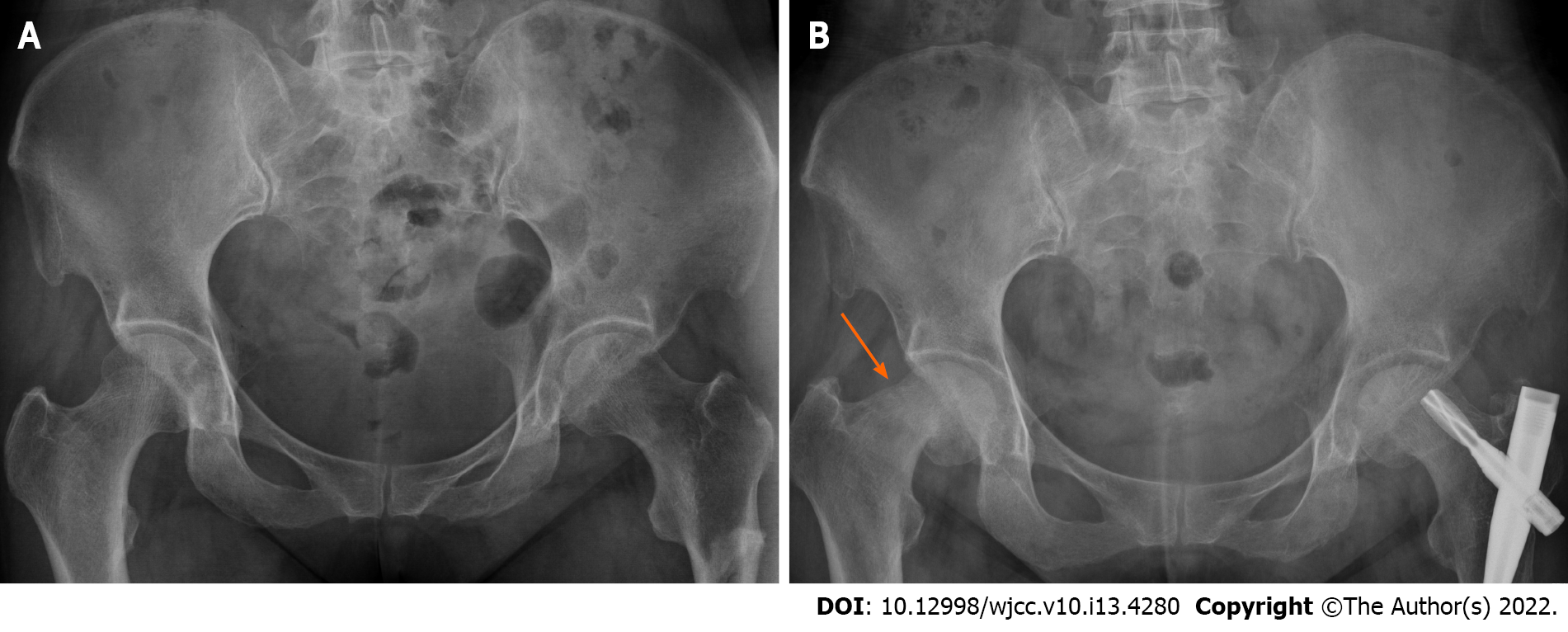
Figure 1 X-ray of the pelvis 1 yr prior to and before surgery.
A: Fracture of the left proximal femur and good continuity of the right femur neck. At that time, the patient had no clinical symptoms in the right hip; B: Fracture line at the upper end of the right femoral neck (orange arrow) and varus deformity of the hip.
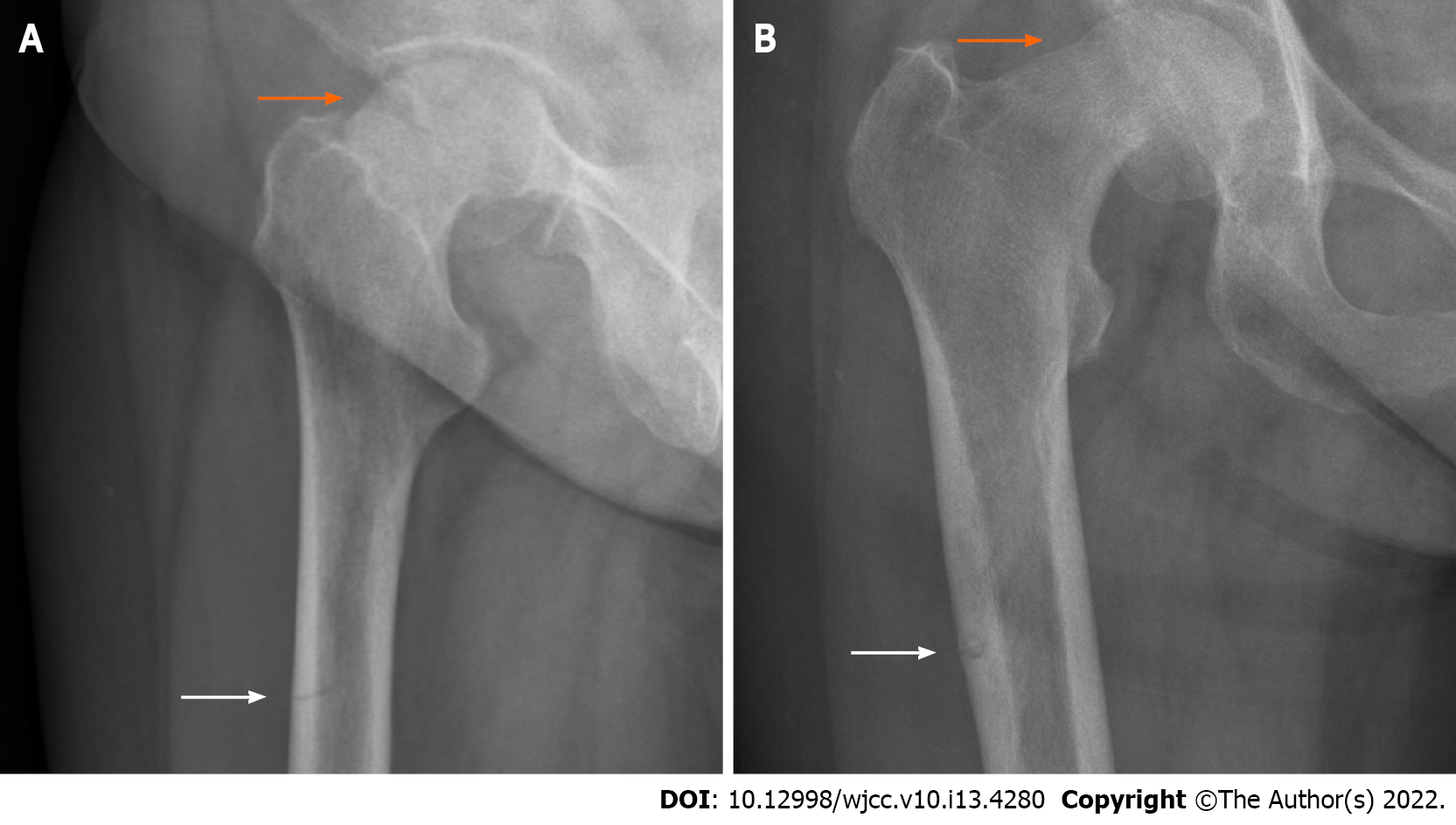
Figure 2 Plain radiograph of the hip joint before an operation.
A: Lateral view; B: Anterior-posterior view. Both show the fracture line at the upper end of the right femoral neck (orange arrow), displacement of the fracture end, varus deformity of the hip, and proximal femur cortical discontinuity (white arrow).
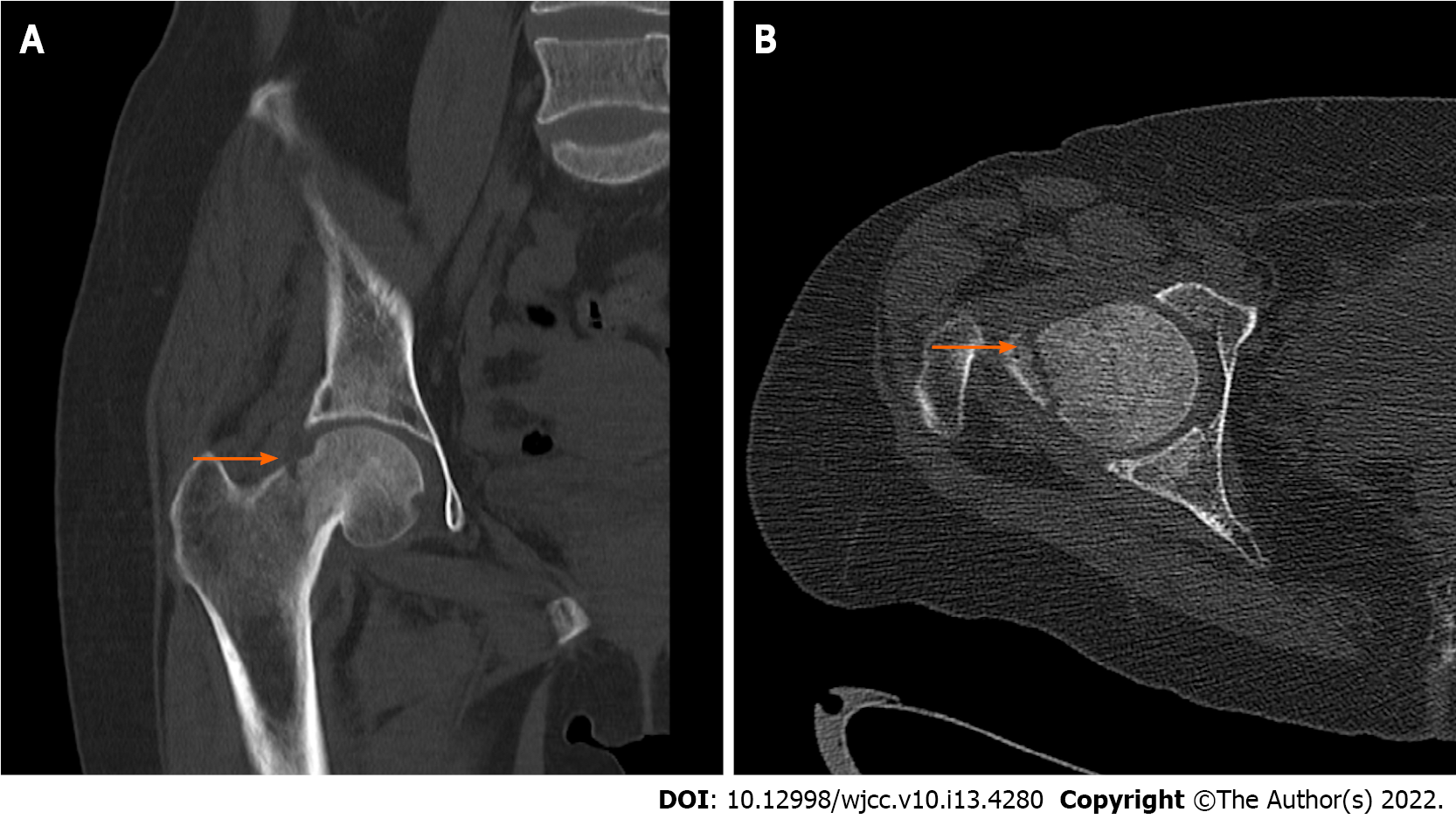
Figure 3 Preoperative computed tomography of the hip joint.
A: Coronal scan; B: Axial scan. Both scans show the fracture line above the femoral neck (arrow), obvious displacement of the fracture end, and varus deformity of the hip.
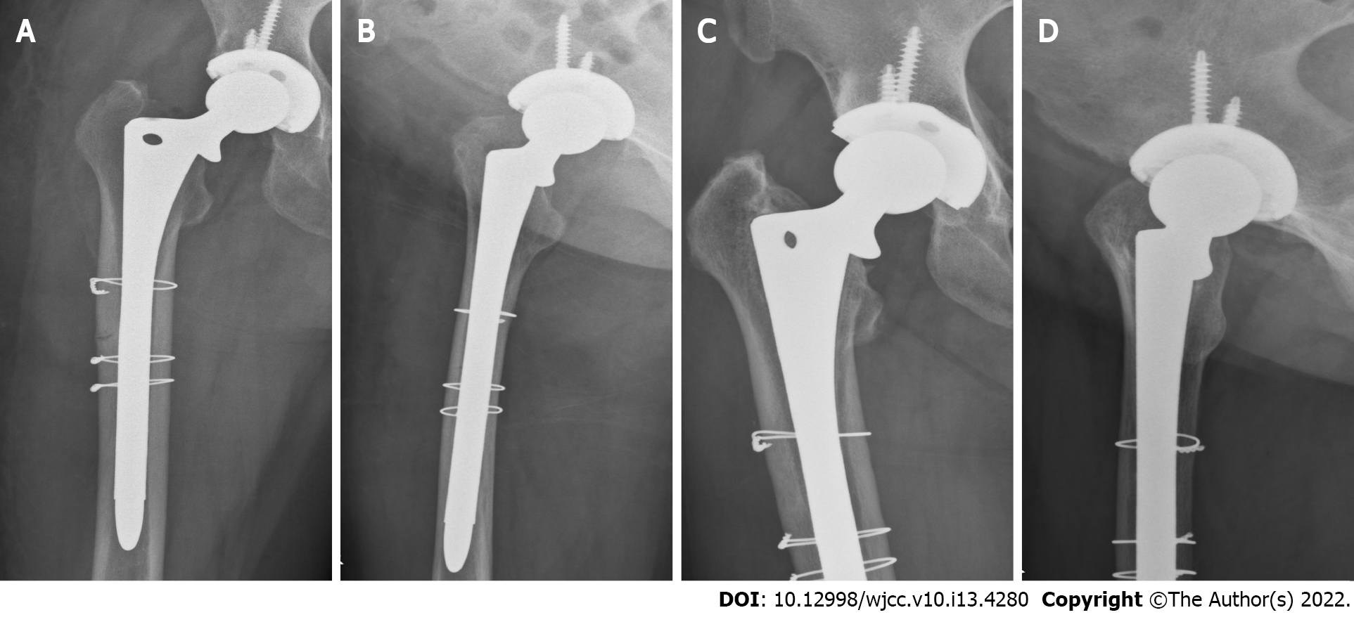
Figure 4 X-rays of hip joint after the operation and 3 mo after the operation.
A: Anterior-posterior view; B: Lateral view. Both views show that, after total hip arthroplasty, the prosthesis was in a good position and the proximal femur was fixed with steel wires to prevent intraoperative fracture; C: Anterior-posterior view; D: Lateral view. Both views show that the right artificial hip prosthesis was in a good position, the fracture line of the right proximal femur disappeared, and the bone fracture healed well.
- Citation: Tang MT, Liu CF, Liu JL, Saijilafu, Wang Z. Multiple stress fractures of unilateral femur: A case report . World J Clin Cases 2022; 10(13): 4280-4287
- URL: https://www.wjgnet.com/2307-8960/full/v10/i13/4280.htm
- DOI: https://dx.doi.org/10.12998/wjcc.v10.i13.4280


