Copyright
©The Author(s) 2022.
World J Clin Cases. May 6, 2022; 10(13): 4264-4272
Published online May 6, 2022. doi: 10.12998/wjcc.v10.i13.4264
Published online May 6, 2022. doi: 10.12998/wjcc.v10.i13.4264
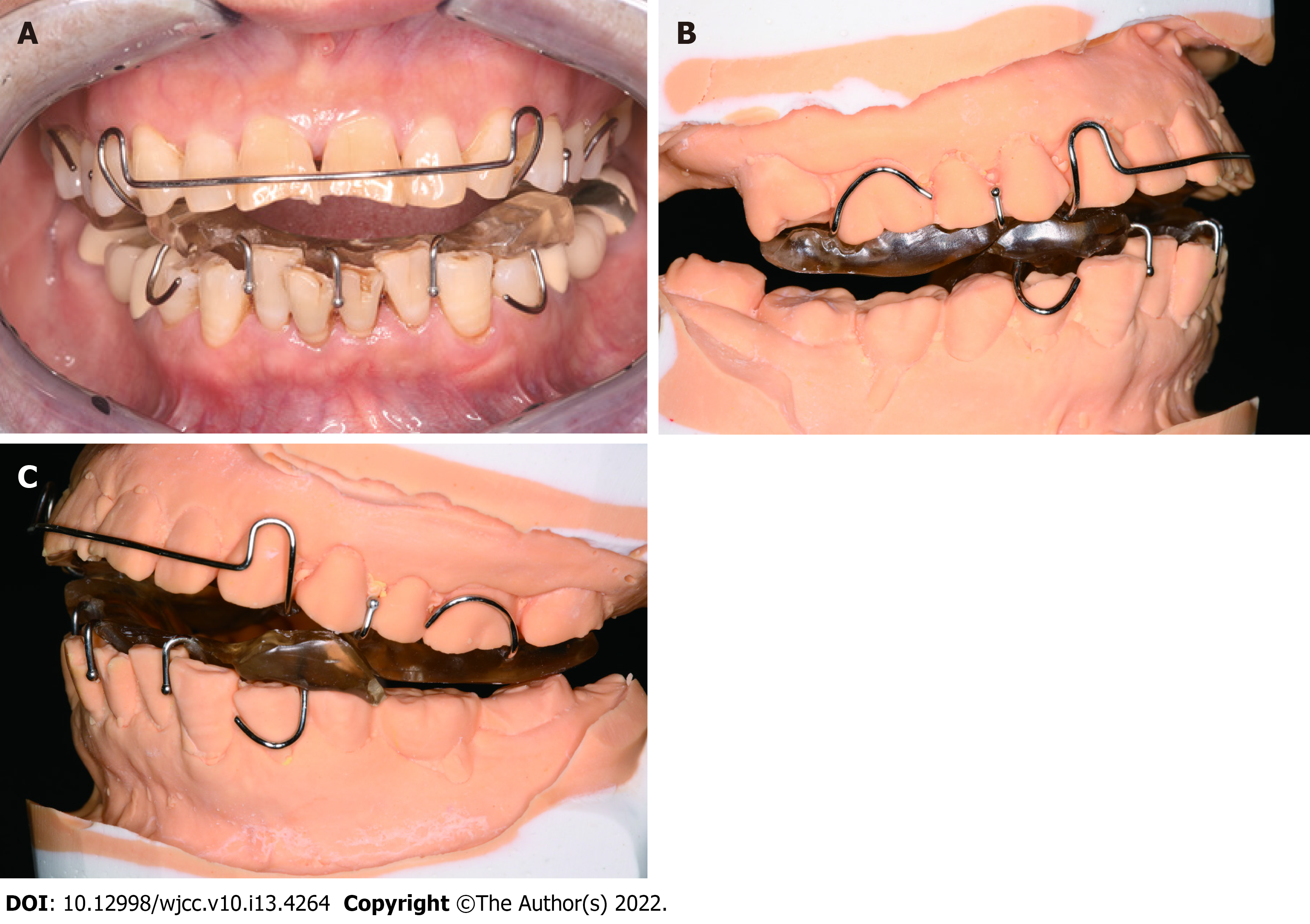
Figure 1 Clinical presentation of the patient wearing the original splint.
A: Intraoral photo of the patient; B: Right side of stone cast; C: Left side of stone cast.
Figure 2 Cone beam computed tomography radiographs of the left temporomandibular joint (March 2017).
A: Sagittal projection; B: Coronal projection.
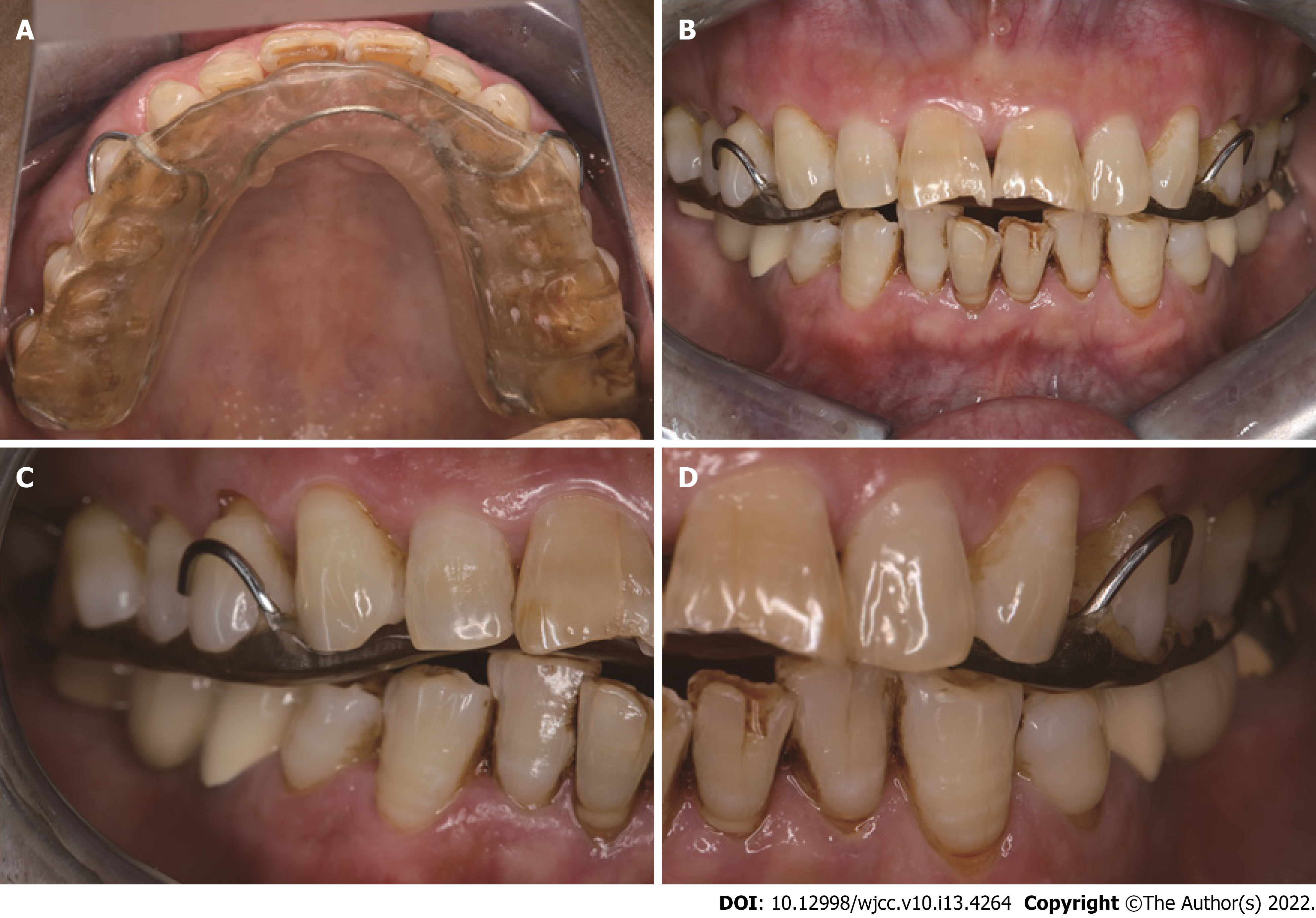
Figure 3 Clinical presentation of wearing the muscle balance occlusal splint.
A: The splint was fixed to the maxillary dentition with clasps; B: Centric occlusion; C: Right posterior occlusion; D: Left posterior occlusion.
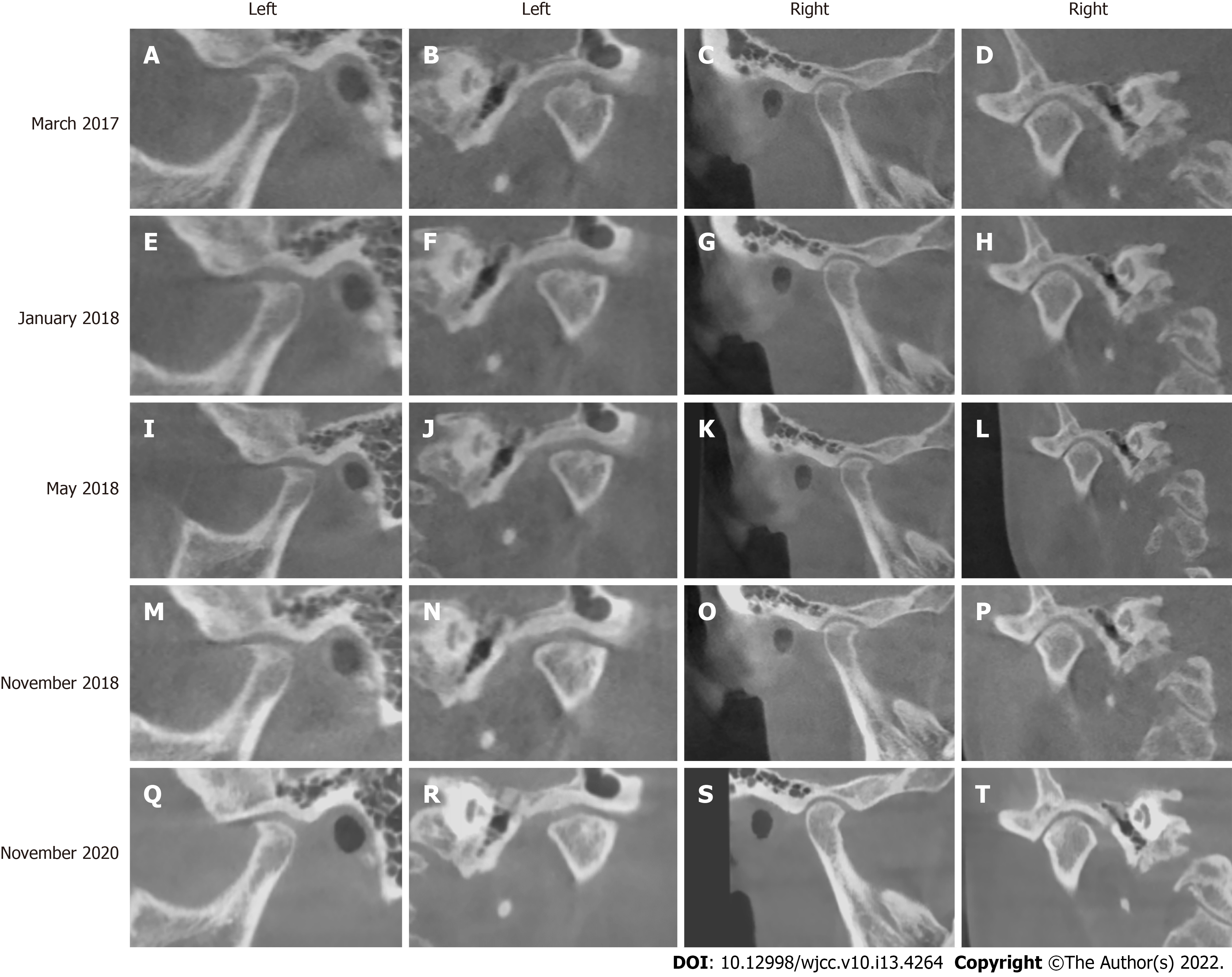
Figure 4 Cone beam computed tomography radiographs of the bilateral temporomandibular joint.
A–D: Computed tomography (CBCT) radiographs obtained in March 2017; E–H: CBCT radiographs obtained in January 2018; I–L: CBCT radiographs obtained in May 2018; M–P: CBCT radiographs in obtained November 2018; Q–T: CBCT radiographs obtained in November 2020.
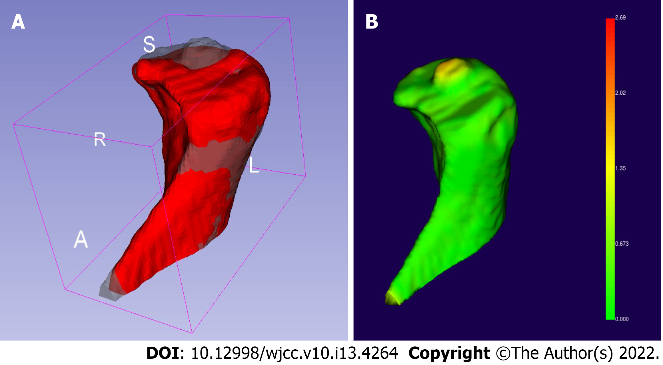
Figure 5 Three-dimensional reconstruction of cone beam computed tomography radiographs.
A: The reconstruction models of the left condyle before treatment (gray model) and 9 mo after treatment (red model) were compared using 3D Slicer version 4.10.2 (https://download.slicer.org); B: We calculated the facial distance of the registration model and found that the 2-mm-high cylindrical osteophyte on top of the condyle had dissolved.
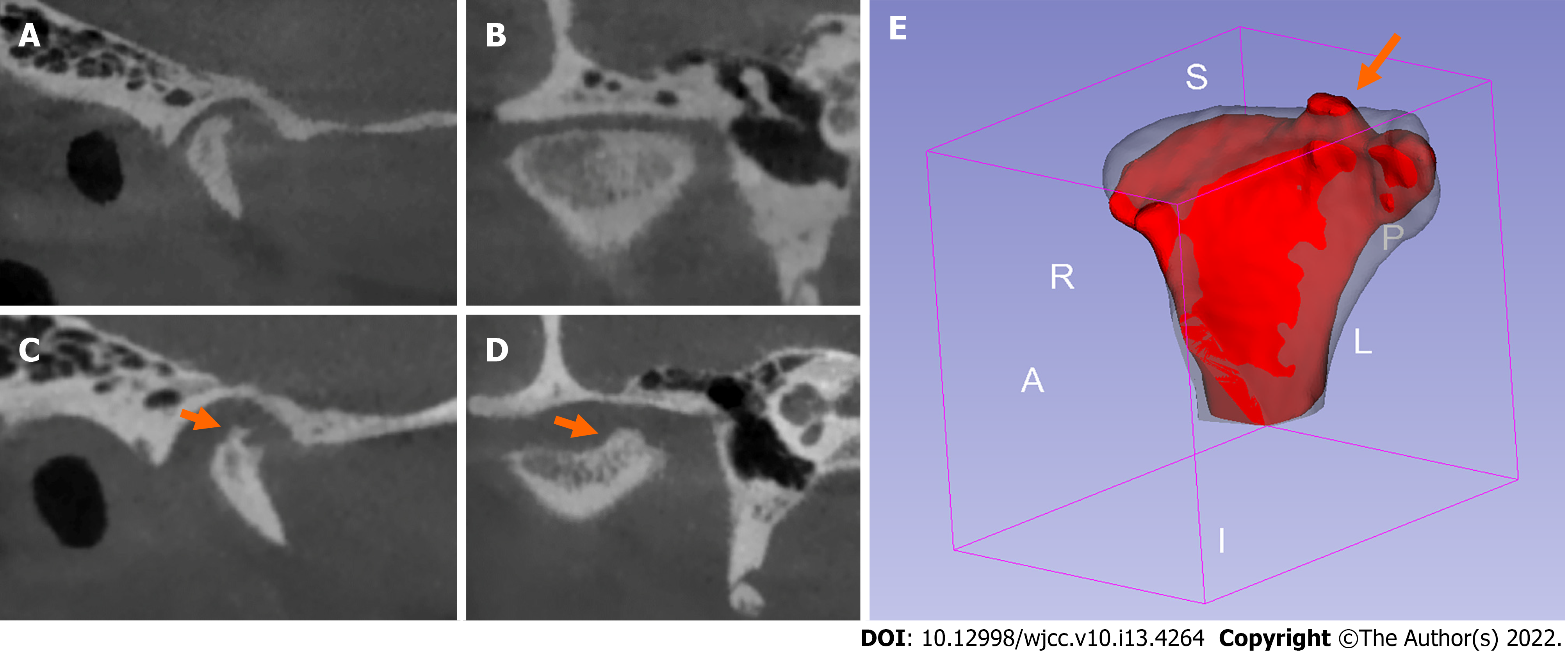
Figure 6 Right condylar changes in a 24-year-old female patient with bilateral condylar resorption before and after treatment with the twin-block occlusal splint.
A: Cone beam computed tomography (CBCT) sagittal radiograph before treatment; B: CBCT coronal radiograph before treatment; C: CBCT sagittal radiograph after treatment; D: CBCT coronal radiograph after treatment; E: Comparison of the condylar three-dimensional model before (gray model) and after (red model) treatment as viewed using 3D Slicer software. Osteophyte (orange arrow) formed on the medial part of the top of the condyle after treatment.
- Citation: Lan KW, Chen JM, Jiang LL, Feng YF, Yan Y. Treatment of condylar osteophyte in temporomandibular joint osteoarthritis with muscle balance occlusal splint and long-term follow-up: A case report. World J Clin Cases 2022; 10(13): 4264-4272
- URL: https://www.wjgnet.com/2307-8960/full/v10/i13/4264.htm
- DOI: https://dx.doi.org/10.12998/wjcc.v10.i13.4264


