Copyright
©The Author(s) 2022.
World J Clin Cases. May 6, 2022; 10(13): 4177-4184
Published online May 6, 2022. doi: 10.12998/wjcc.v10.i13.4177
Published online May 6, 2022. doi: 10.12998/wjcc.v10.i13.4177
Figure 1 Magnetic resonance image with balanced turbo field echo, showing that the pregnant uterus vertically stretched the bladder (arrow).
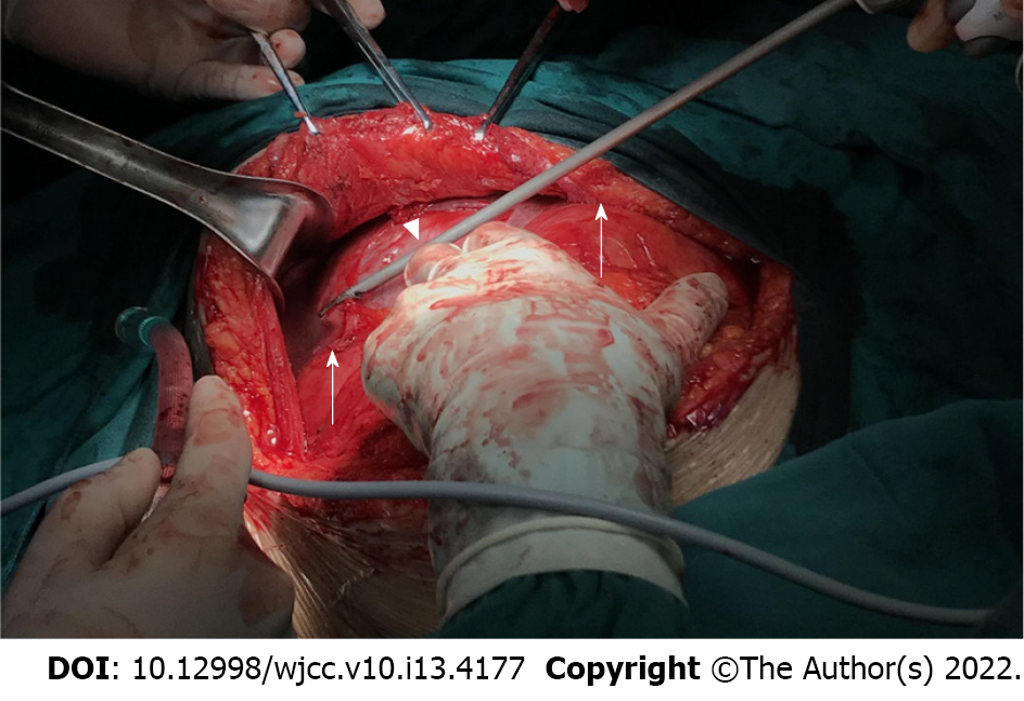
Figure 2 Left lateral traction of the neobladder (arrow) after sharp dissection of the adhesion between the uterus (arrow head) and neobladder with an ultrasonic scalpel.
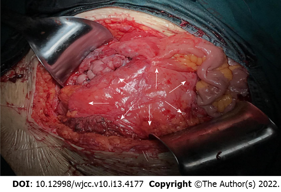
Figure 3 Right lateral longitudinal incision (arrow) and neobladder (arrow).
- Citation: Ruan J, Zhang L, Duan MF, Luo DY. Pregnancy and delivery after augmentation cystoplasty: A case report and review of literature. World J Clin Cases 2022; 10(13): 4177-4184
- URL: https://www.wjgnet.com/2307-8960/full/v10/i13/4177.htm
- DOI: https://dx.doi.org/10.12998/wjcc.v10.i13.4177


