Copyright
©The Author(s) 2022.
World J Clin Cases. May 6, 2022; 10(13): 4161-4170
Published online May 6, 2022. doi: 10.12998/wjcc.v10.i13.4161
Published online May 6, 2022. doi: 10.12998/wjcc.v10.i13.4161
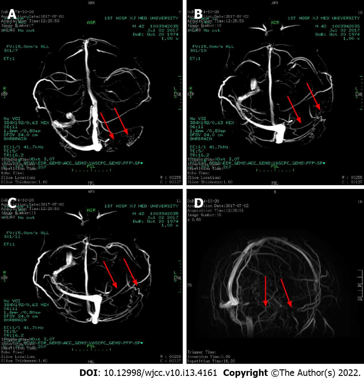
Figure 1 Magnetic resonance venography and digital subtraction angiography images.
A and B: The left sigmoid sinus and the left transverse sinus are not obviously visualized, nor are the right and right venous ends of the superior sagittal sinus; C and D: The abnormal signal in the left sigmoid sinus and transverse sinus and abnormal enhancement indicate venous sinusitis and thrombosis.
Figure 2 Hematoxylin and eosin staining of bone marrow.
Scale bars = 80 µm; × 200 magnification. The arrows show immune cells. A and B: Hematoxylin and eosin staining shows that bone marrow hematopoietic cell hyperplasia is significantly active, erythroid hyperplasia is significantly active, and granulocyte hyperplasia is reduced; C and D: Net dyeing showing that fibrous tissue hyperplasia in focal areas between trabecular bones is obvious.
Figure 3 Thrombosis gene test: SERPINC1 (NM-000488).
- Citation: Wufuer G, Wufuer K, Ba T, Cui T, Tao L, Fu L, Mao M, Duan MH. Primary myelofibrosis with thrombophilia as first symptom combined with thalassemia and Gilbert syndrome: A case report. World J Clin Cases 2022; 10(13): 4161-4170
- URL: https://www.wjgnet.com/2307-8960/full/v10/i13/4161.htm
- DOI: https://dx.doi.org/10.12998/wjcc.v10.i13.4161


