Copyright
©The Author(s) 2022.
World J Clin Cases. Apr 26, 2022; 10(12): 3959-3965
Published online Apr 26, 2022. doi: 10.12998/wjcc.v10.i12.3959
Published online Apr 26, 2022. doi: 10.12998/wjcc.v10.i12.3959
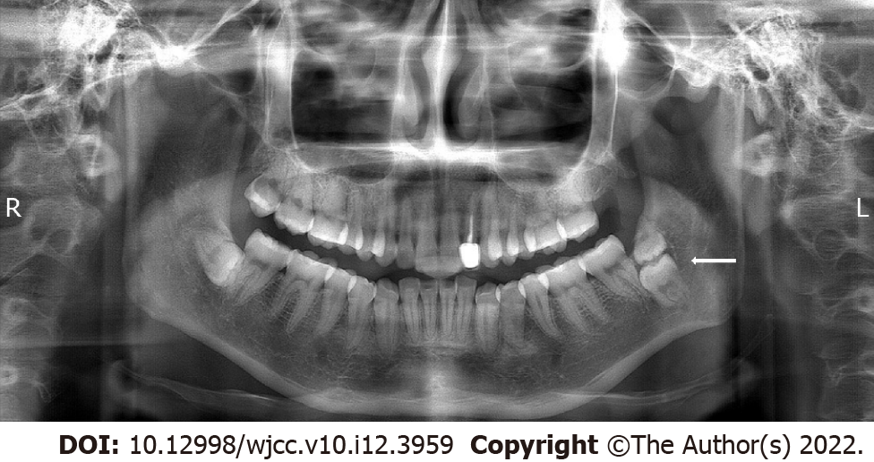
Figure 1 Teeth #17 and #67 in the vertical direction, showing impacted kissing molars.
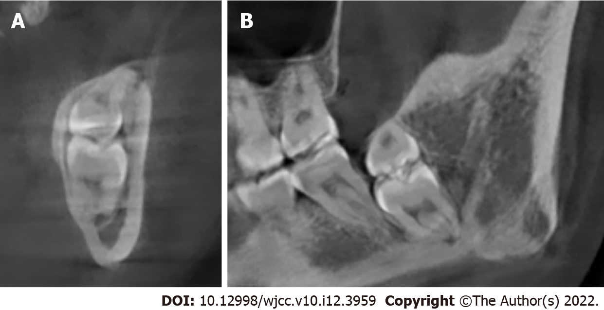
Figure 2 Cone-beam computed tomography image of kissing molars, the root of tooth #17 appears close to the inferior alveolar nerve, the kissing molars and tooth #18 were close, and the distal bone of tooth #18 was absorbed.
A: Coronal plane; B: Sagittal plane.
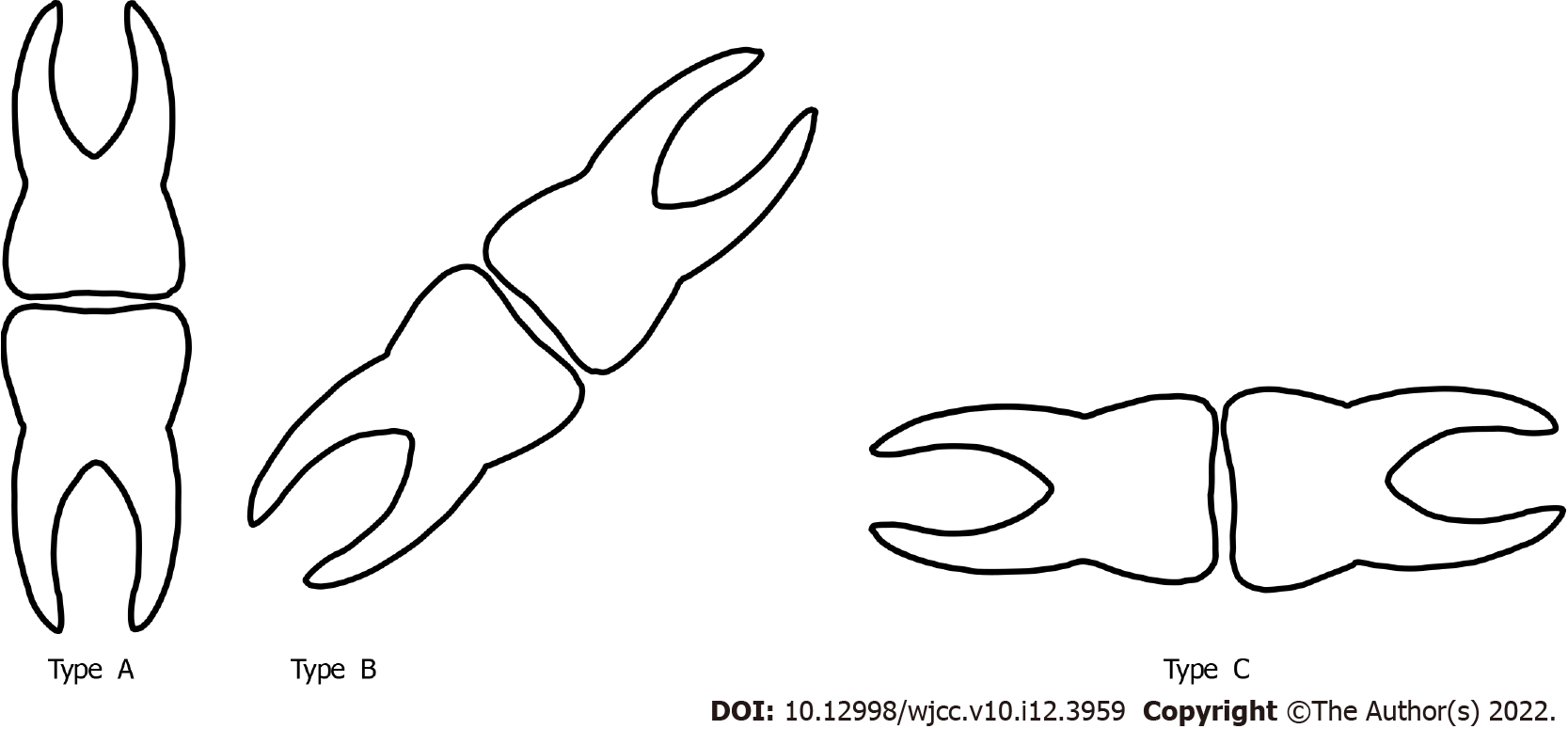
Figure 3 Classification of kissing molars according to the impaction direction (from left to right: Type A, vertical, Type B, tilted, Type C, transverse).
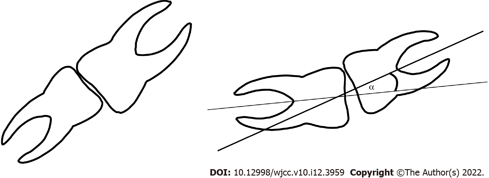
Figure 4 Kissing molars; the overlapping region of the two teeth, and the acute angle formed by the long axis of the two teeth should be considered when identifying kissing molars.
- Citation: Wen C, Jiang R, Zhang ZQ, Lei B, Yan YZ, Zhong YQ, Tang L. Vertical direction impaction of kissing molars: A case report . World J Clin Cases 2022; 10(12): 3959-3965
- URL: https://www.wjgnet.com/2307-8960/full/v10/i12/3959.htm
- DOI: https://dx.doi.org/10.12998/wjcc.v10.i12.3959


