Copyright
©The Author(s) 2022.
World J Clin Cases. Apr 26, 2022; 10(12): 3944-3950
Published online Apr 26, 2022. doi: 10.12998/wjcc.v10.i12.3944
Published online Apr 26, 2022. doi: 10.12998/wjcc.v10.i12.3944
Figure 1 Ultrasonography identified a locally discontinuous band of strong echo in the abdominal wall of the right inguinal area.
An inhomogeneous echo mass (dimensions: 3.9 cm ×1.5 cm) was detected on its deep surface.
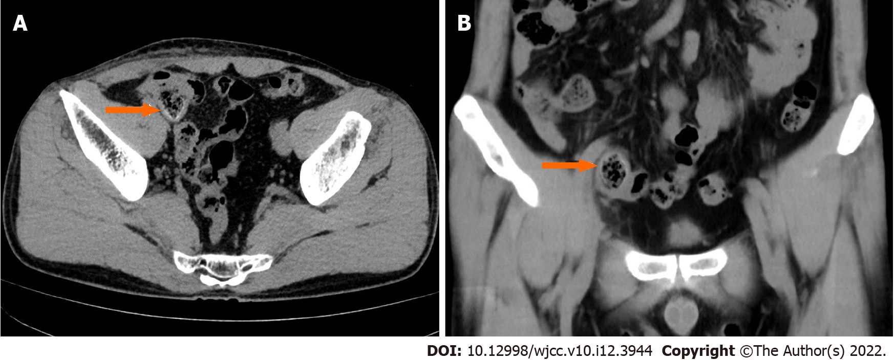
Figure 2 Computed tomography revealed a circinate high-density image with short segmental thickening of the ileum stuck to the abdominal wall (orange arrow) with no evidence of recurrent inguinal hernia.
Figure 3 During surgery.
A: Adhesion of the ileum loop to the right inguinal abdominal wall; B: Migration of the polypropylene mesh plug (MP) into the intra-peritoneal cavity; C: The internal ring was 1.0 cm in diameter, but no hernia sac was found; D: Specimen examination revealed erosion of the iliac wall due to the MP.
Figure 4 Postoperative pathology showed chronic inflammatory changes in the small intestine mucosa, with focal granulomatous tissue formation, and focal abscess in the serosa.
- Citation: Xie TH, Wang Q, Ha SN, Cheng SJ, Niu Z, Ren XX, Sun Q, Jin XS. Mesh plug erosion into the small intestine after inguinal hernia repair: A case report . World J Clin Cases 2022; 10(12): 3944-3950
- URL: https://www.wjgnet.com/2307-8960/full/v10/i12/3944.htm
- DOI: https://dx.doi.org/10.12998/wjcc.v10.i12.3944


