Copyright
©The Author(s) 2022.
World J Clin Cases. Apr 26, 2022; 10(12): 3936-3943
Published online Apr 26, 2022. doi: 10.12998/wjcc.v10.i12.3936
Published online Apr 26, 2022. doi: 10.12998/wjcc.v10.i12.3936
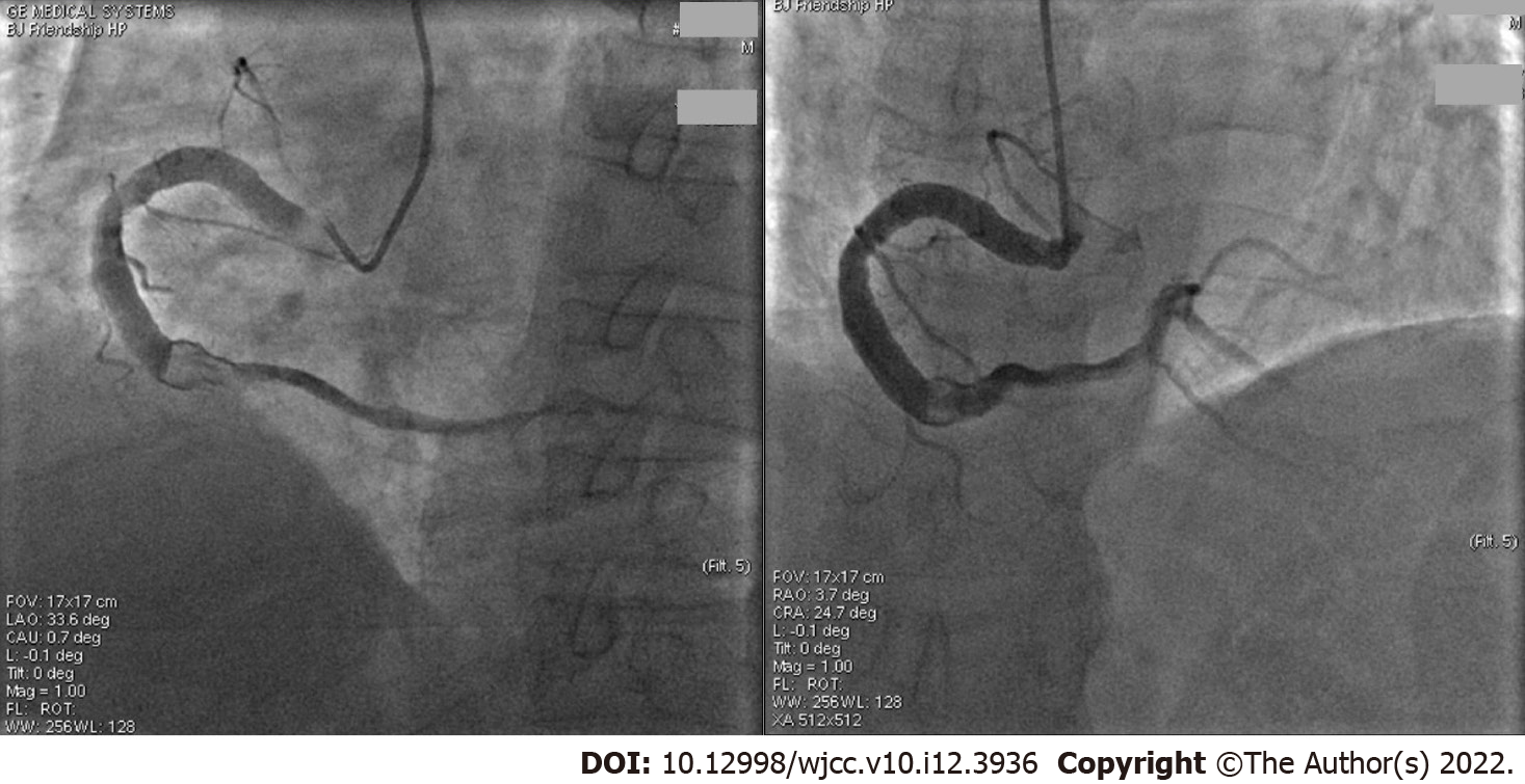
Figure 1 The first coronary angiography.
This angiography was performed 3 d after admission to hospital with chest pain (video), Q wave on electrocardiogram, and increased myocardial injury markers. The two images show aneurysmal dilation in the entire right coronary artery (RCA) with a diameter of 6.60 mm, four fold of a 5 French catheter and more than 1.70 fold of the normal RCA. The middle of the RCA presented with 70%-80% limited stenosis, showed a thrombus shadow after the second turning point, and the forward blood flow was thrombolysis in myocardial infarction grade 3. No additional percutaneous coronary intervention (PCI) was performed after a discussion with the PCI group because the symptoms were relieved and the coronary blood flow was unobstructed.
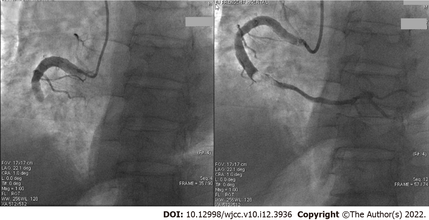
Figure 2 The second coronary angiography.
This angiography was performed in the second month (video), one month after treatment with a combination of aspirin (100 mg once per day) and clopidogrel (75 mg/d), as well as low-molecular-weight heparin. The thrombus in the right coronary artery disappeared and forward blood flow was thrombolysis in myocardial infarction grade 3. The aspirin and clopidogrel were continued.
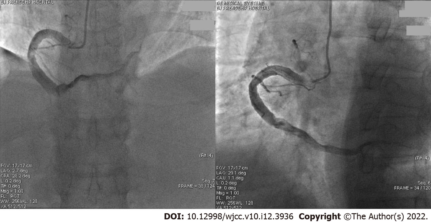
Figure 3 The third coronary angiography.
This angiography was performed in the fifth month after sudden chest pain (video). The left panel shows that the middle of the right coronary artery (RCA) was 100% occluded and a thrombus shadow could be seen. Thrombus aspiration was performed immediately from the middle to distant RCA, and a small amount of white flocculent thrombus was extracted. The right panel shows reperfusion of the RCA, which relieved the patient's chest pain. The antithrombotic agents were changed to aspirin (100 mg/d) and ticagrelor (ticagrelor tablet, 90 mg twice per day) with 1 wk of LMWH.
Figure 4 The fourth coronary angiography.
This angiography was performed on the sixth month because of slight chest tightness that occurred without obvious electrocardiogram changes (video). The coronary angiography showed the middle of the right coronary artery was slightly blurred. Chest tightness was considered a side effect of the ticagrelor. Thus, the antithrombotic agents were not changed.
- Citation: Liu RF, Gao XY, Liang SW, Zhao HQ. Antithrombotic treatment strategy for patients with coronary artery ectasia and acute myocardial infarction: A case report. World J Clin Cases 2022; 10(12): 3936-3943
- URL: https://www.wjgnet.com/2307-8960/full/v10/i12/3936.htm
- DOI: https://dx.doi.org/10.12998/wjcc.v10.i12.3936


