Copyright
©The Author(s) 2022.
World J Clin Cases. Apr 26, 2022; 10(12): 3899-3906
Published online Apr 26, 2022. doi: 10.12998/wjcc.v10.i12.3899
Published online Apr 26, 2022. doi: 10.12998/wjcc.v10.i12.3899
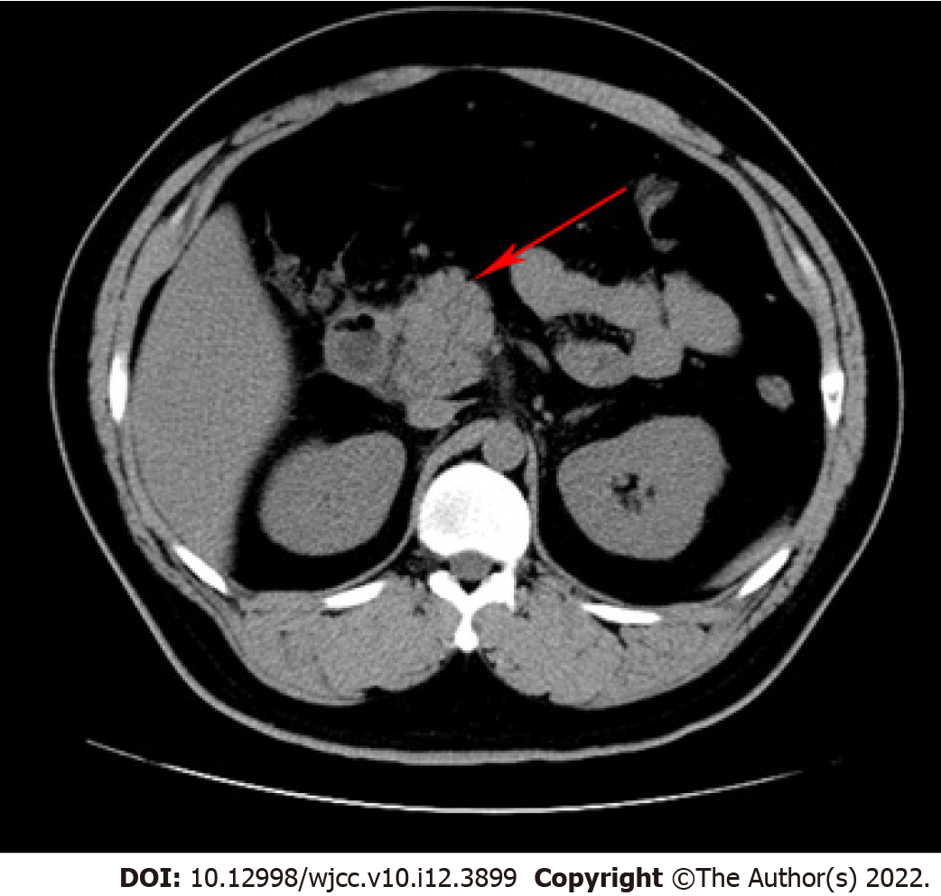
Figure 1 Computed tomography of the abdomen revealed pancreatitis (red arrow).
Figure 2 An electrocardiogram.
The electrocardiogram revealed sinus tachycardia with a heart rate of 145 beats per minute.
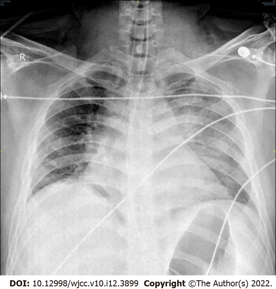
Figure 3 Chest X-ray.
The chest X-ray revealed exudative changes in the left lung.
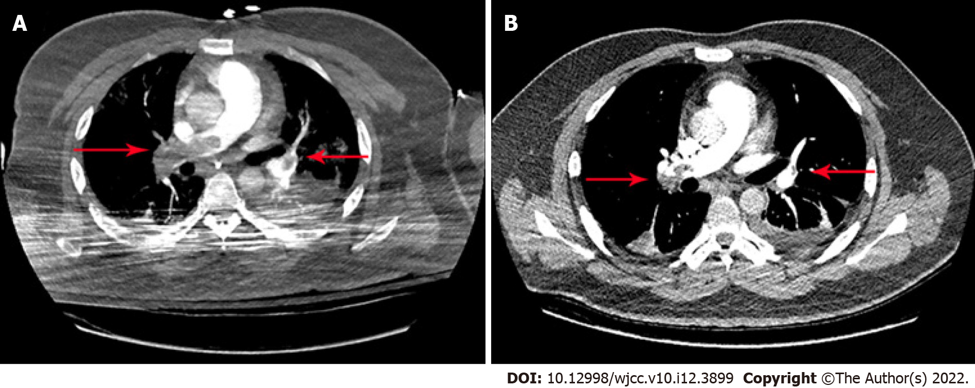
Figure 4 Computed tomography angiography findings.
A: Computed tomography angiography of the chest revealed pulmonary embolus (the right main pulmonary artery and multiple lobar pulmonary arteries) (red arrow); B: Partial resolution of the thrombosis was documented on follow-up chest computed tomography angiography (red arrow).
- Citation: Yan LL, Jin XX, Yan XD, Peng JB, Li ZY, He BL. Combined use of extracorporeal membrane oxygenation with interventional surgery for acute pancreatitis with pulmonary embolism: A case report. World J Clin Cases 2022; 10(12): 3899-3906
- URL: https://www.wjgnet.com/2307-8960/full/v10/i12/3899.htm
- DOI: https://dx.doi.org/10.12998/wjcc.v10.i12.3899


