Copyright
©The Author(s) 2022.
World J Clin Cases. Apr 26, 2022; 10(12): 3879-3885
Published online Apr 26, 2022. doi: 10.12998/wjcc.v10.i12.3879
Published online Apr 26, 2022. doi: 10.12998/wjcc.v10.i12.3879
Figure 1 Preoperative radiological findings of the right knee.
A, B: Plain anteroposterior and lateral radiographs revealing a huge avulsion fracture of the intercondylar eminence of the tibia containing the attachment of site of both the anterior and posterior cruciate ligaments; C, D: Coronal and sagittal computed tomography scans showing the fracture line that reached the medial tibial plateau (arrow); E–G: Coronal and sagittal T2-weighted magnetic resonance imaging of the right knee indicating the medial collateral ligament tear (arrow), and tear of both the medial and lateral menisci (arrowheads).
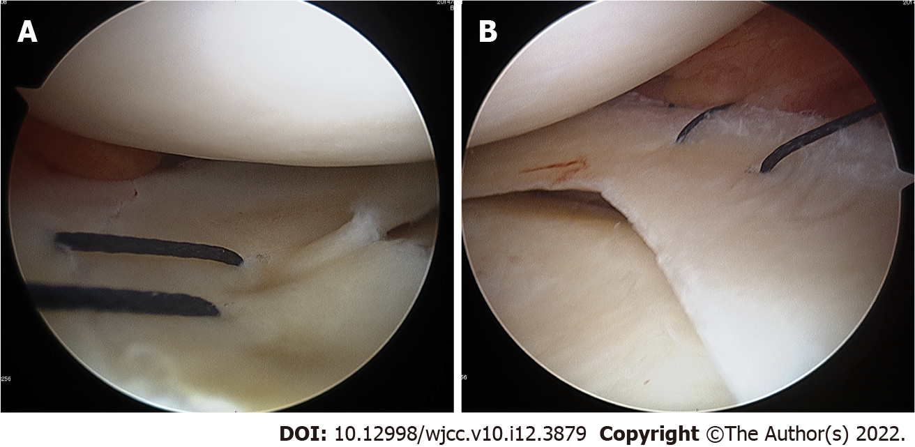
Figure 2 Intraoperative arthroscopic findings of the lateral and medial menisci.
A: The lateral meniscus tear was sutured with the inside-out technique; B: The medial meniscus tear was sutured under direct view.
Figure 3 Postoperative plain radiographs of the right knee.
A, B: Plain anteroposterior and lateral radiographs revealing satisfactory reduction and fixation.
Figure 4 Follow-up imaging at 1 yr post-surgery.
A, B: Coronal and sagittal computed tomography scans of the right knee show bone union with excellent alignment; C, D: Valgus stress radiographs of the bilateral knee joints showed no lateral instability of the right knee.
Figure 5 Intraoperative arthroscopic findings at 1 yr post-surgery.
A: The tension of the anterior cruciate ligament is moderate; B, C: Both the lateral and medial menisci show complete healing.
- Citation: Yoshida K, Hakozaki M, Kobayashi H, Kimura M, Konno S. Surgical treatment for a combined anterior cruciate ligament and posterior cruciate ligament avulsion fracture: A case report. World J Clin Cases 2022; 10(12): 3879-3885
- URL: https://www.wjgnet.com/2307-8960/full/v10/i12/3879.htm
- DOI: https://dx.doi.org/10.12998/wjcc.v10.i12.3879


