Copyright
©The Author(s) 2022.
World J Clin Cases. Apr 26, 2022; 10(12): 3866-3871
Published online Apr 26, 2022. doi: 10.12998/wjcc.v10.i12.3866
Published online Apr 26, 2022. doi: 10.12998/wjcc.v10.i12.3866
Figure 1 Results of the computed tomography of the abdomen showing edema and bowel wall thickening with hypodensity in the sigmoid colon and descending colon.
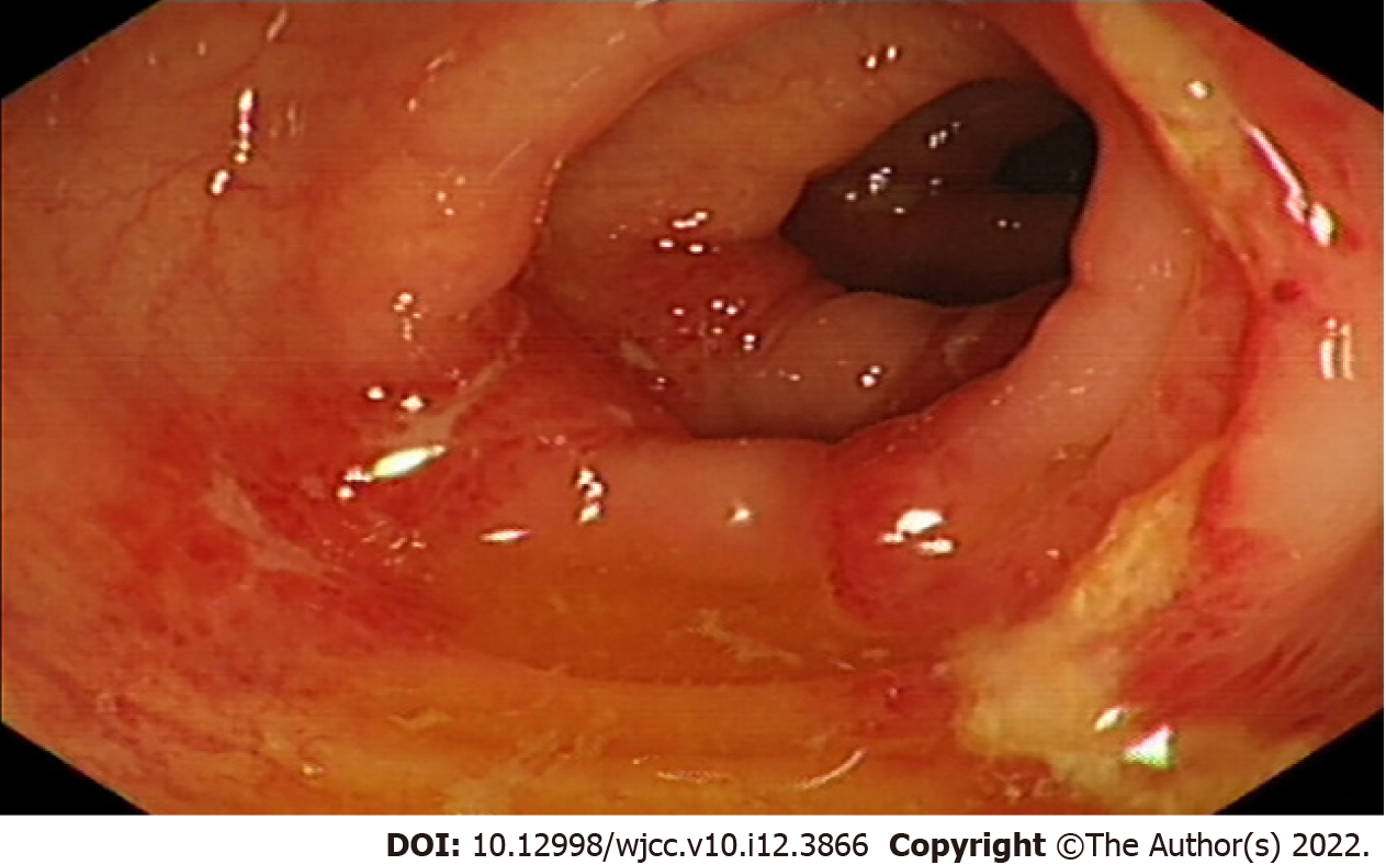
Figure 2 Results of colonoscopy illustrating hyperemia, edema and erosion of the mucosa with superficial ulceration and a yellow-white coating located at the descending colon and sigmoid colon.
- Citation: Cui MH, Hou XL, Liu JY. Ischemic colitis after receiving the second dose of a COVID-19 inactivated vaccine: A case report. World J Clin Cases 2022; 10(12): 3866-3871
- URL: https://www.wjgnet.com/2307-8960/full/v10/i12/3866.htm
- DOI: https://dx.doi.org/10.12998/wjcc.v10.i12.3866


