Copyright
©The Author(s) 2022.
World J Clin Cases. Apr 26, 2022; 10(12): 3834-3841
Published online Apr 26, 2022. doi: 10.12998/wjcc.v10.i12.3834
Published online Apr 26, 2022. doi: 10.12998/wjcc.v10.i12.3834
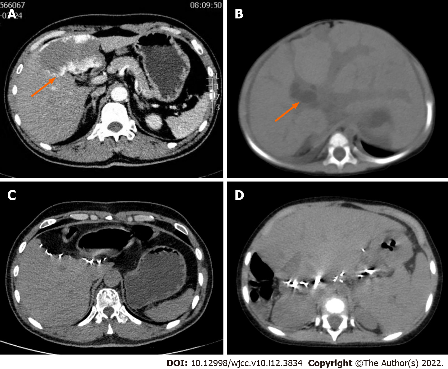
Figure 1 Preoperative contrast-enhanced abdominal computed tomography and postoperative abdominal computed tomography of the donor and recipient liver.
A: Hemangioma was indicated by orange arrow; B: Dilated intrahepatic bile duct showed by orange arrow, and diffuse hepatomegaly could be seen; C: The liver of the donor was regenerated half a year after the operation without hemangioma recurrence; D: The liver allograft transplanted to the recipient was regenerated 2 years after the operation without hemangioma recurrence.
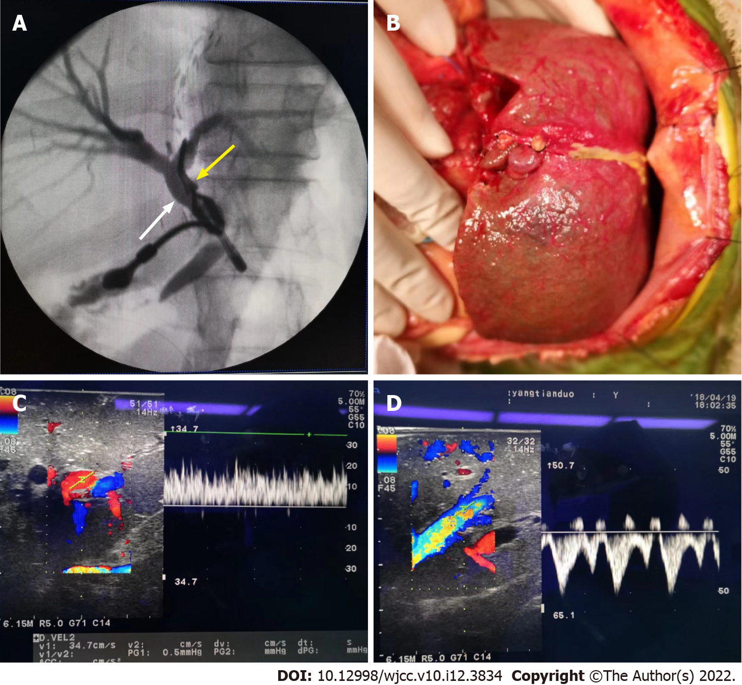
Figure 2 Intraoperative intrahepatic cholangiography and reperfusion of the liver allograft, postoperative blood flow of portal vein and hepatic vein.
A: Intraoperative intrahepatic cholangiography indicate the common bile duct (white arrow) and left hepatic bile duct was clipped, and no variation was observed; B: Intraoperative liver allograft reperfusion; C: Portal vein blood flow after reperfusion was 22 cm/s; D: Hepatic venous blood flow after reperfusion was satisfactory without constriction.
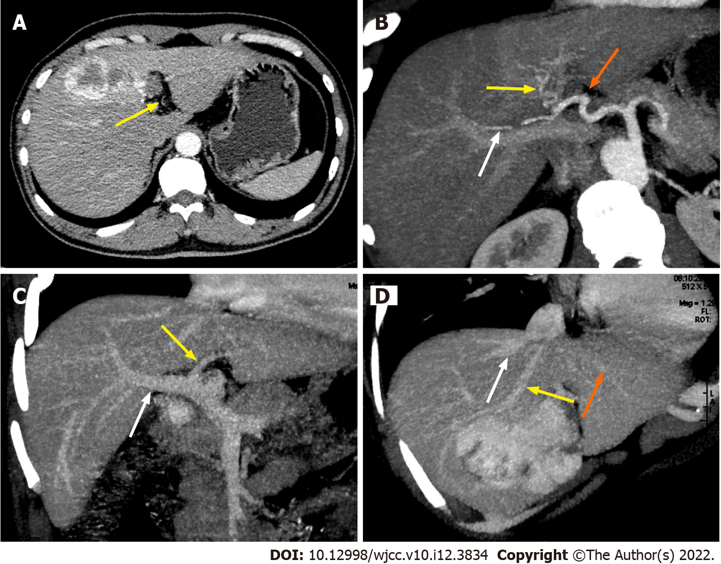
Figure 3 Preoperative contrast-enhanced abdominal computed tomography and reconstruction of the donor liver allograft vessels.
A: Yellow arrow indicate the left hepatic artery supplying the segment 2 and 3 of the liver which is not big enough to be clearly seen in reconstruction figure; B: Hepatic artery reconstruction of the liver allograft (white arrow indicate right hepatic artery supplying right lobe of the liver; yellow arrow indicate middle hepatic artery supplying segment 4 of the liver; orange arrows indicate the left hepatic artery supplying the segment 2 and 3 of the liver); C: Portal vein reconstruction of the liver allograft (white arrow indicate right portal vein; yellow arrow indicate left portal vein); D: Hepatic vein reconstruction of the live allograft (white arrow indicate right hepatic vein; yellow arrow indicate middle hepatic vein; orange arrows indicate the left hepatic vein).
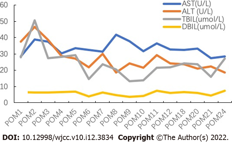
Figure 4 Course of alanine aminotransferase, aspartate aminotransferase, total bilirubin, and direct bilirubin during the perioperative period.
AST: Aspartate aminotransferase; ALT: Alanine aminotransferase; DBIL: Direct bilirubin; POM: Postoperative month; TBIL: Total bilirubin.
- Citation: Li SX, Tang HN, Lv GY, Chen X. Pediatric living donor liver transplantation using liver allograft after ex vivo backtable resection of hemangioma: A case report. World J Clin Cases 2022; 10(12): 3834-3841
- URL: https://www.wjgnet.com/2307-8960/full/v10/i12/3834.htm
- DOI: https://dx.doi.org/10.12998/wjcc.v10.i12.3834


