Copyright
©The Author(s) 2022.
World J Clin Cases. Apr 26, 2022; 10(12): 3814-3821
Published online Apr 26, 2022. doi: 10.12998/wjcc.v10.i12.3814
Published online Apr 26, 2022. doi: 10.12998/wjcc.v10.i12.3814
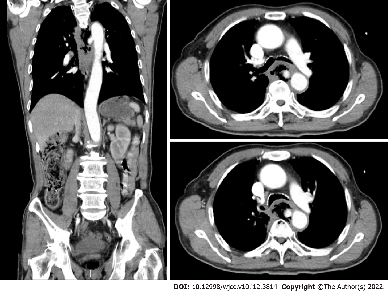
Figure 1 computer tomography scan.
The descending aortic pseudoaneurysm broke into the esophagus.
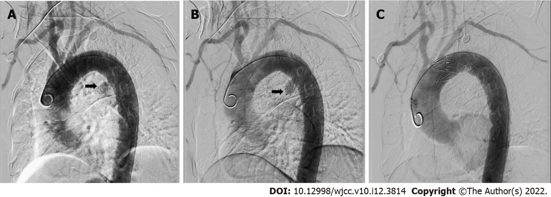
Figure 2 Aortic angiography.
A: There was leakage of contrast in the initial segment of the descending aorta; B: The pseudoaneurysm of the descending aorta; C: The pseudoaneurysm was blocked, and the bleeding was controlled after thoracic endovascular aortic repair.
- Citation: Zhong XQ, Li GX. Successful management of life-threatening aortoesophageal fistula: A case report and review of the literature. World J Clin Cases 2022; 10(12): 3814-3821
- URL: https://www.wjgnet.com/2307-8960/full/v10/i12/3814.htm
- DOI: https://dx.doi.org/10.12998/wjcc.v10.i12.3814


