Copyright
©The Author(s) 2022.
World J Clin Cases. Apr 6, 2022; 10(10): 3251-3260
Published online Apr 6, 2022. doi: 10.12998/wjcc.v10.i10.3251
Published online Apr 6, 2022. doi: 10.12998/wjcc.v10.i10.3251
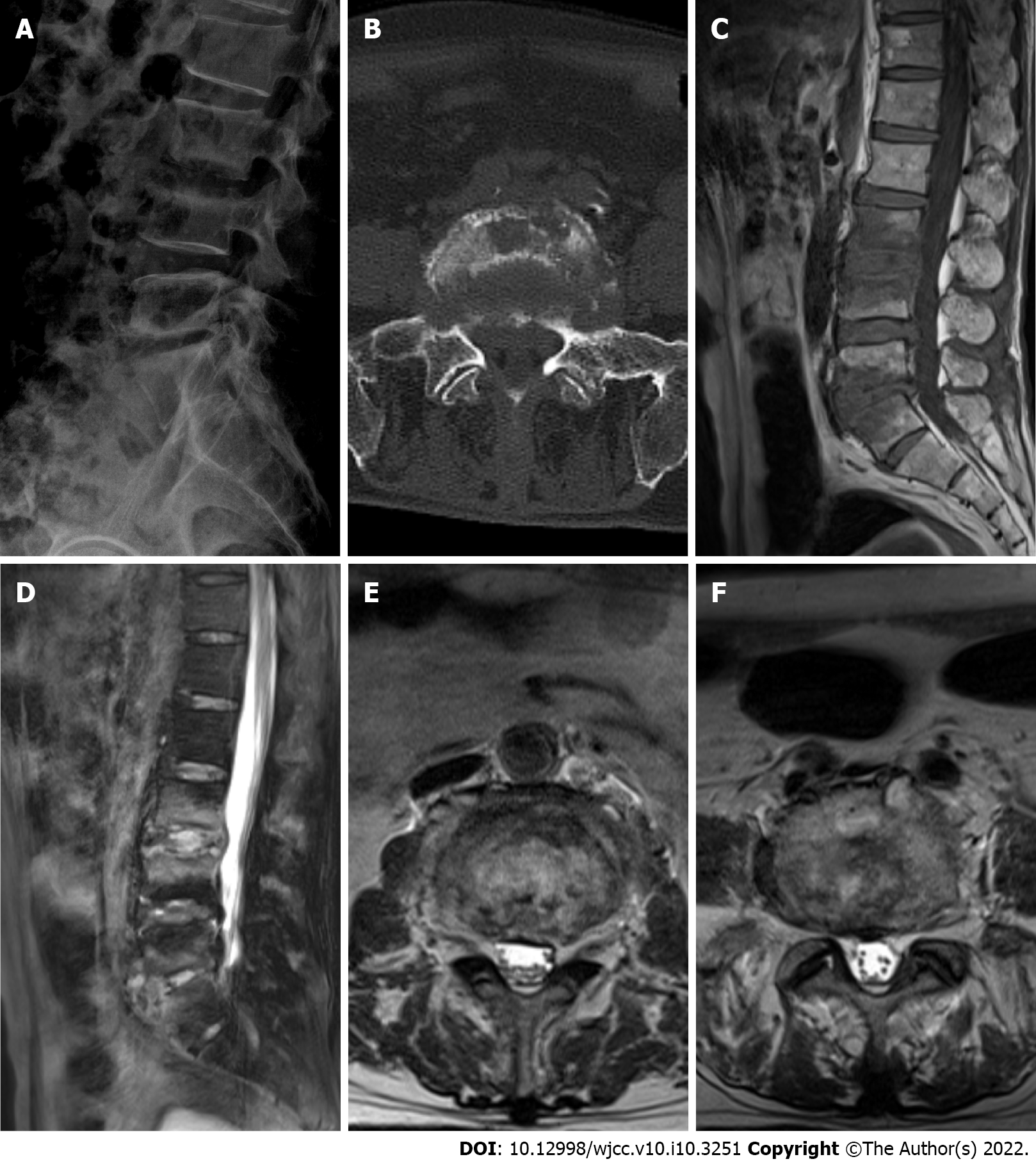
Figure 1 Medical imaging examinations before therapy.
A: X-ray image showing increased heterogeneity of bone density at the edge of the third lumbar (L3) vertebra to the first sacral (S1) vertebra. The fifth lumbar (L5) vertebra and S1 bodies had marginal hyperostosis with incomplete posterior border continuity, the L5 body was displaced slightly posteriorly, and the L5/S1 intervertebral space was narrowed; B: Axial computed tomography image showing bone destruction and hyperplasia at the edges of the L5 and S1 bodies, a small amount of low-density shadow encircling the paravertebral space, and bone destruction at the right sacroiliac joint surface; C and D: Sagittal T1WI and T2WI magnetic resonance imaging (MRI) showing abnormal bone signal at the margins of L3 and the fourth lumbar (L4), and L5 and S1, with a long T1 and mixed long T2 signal; E and F: Axial T2WI in MRI showing a decreased signal at the L3/L4 disc (E), L5/S1 disc (F), rim of the visible soft tissue shadow of the annular bulge, and slight narrowing of the spinal canal at the corresponding level.
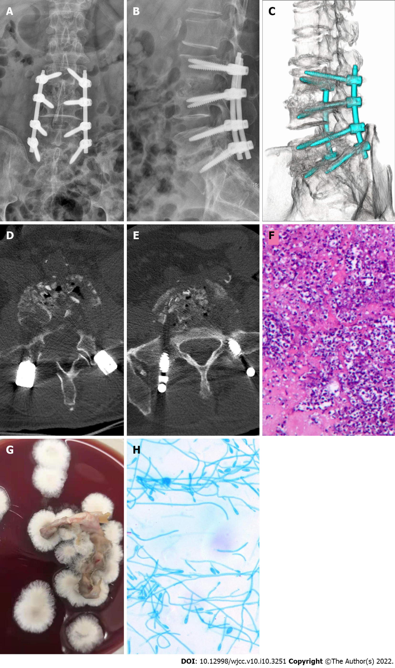
Figure 2 Medical imaging, pathology and microbiology examination after surgery.
A and B: X-ray image showing that the lumbar internal fixation device was in a good position; C-E: Three-dimensional computed tomography image showing sufficient bone grafting in the L3/L4 and L5/S1 intervertebral spaces; F: Pathological examination results showing a large number of inflammatory cells in the tissues examined, and the staining revealed PAS (+), and acid resistance (-); G: Lesion tissue culture day 7 (blood agar medium, 30 °C, 7 d) showed that colonies were cashmere-like and the back was gray-black; H: Under the microscope (lactic acid phenol cotton blue staining, × 400), most of the hyphae were irregularly branched, producing round or oval lateral and terminal conidia.
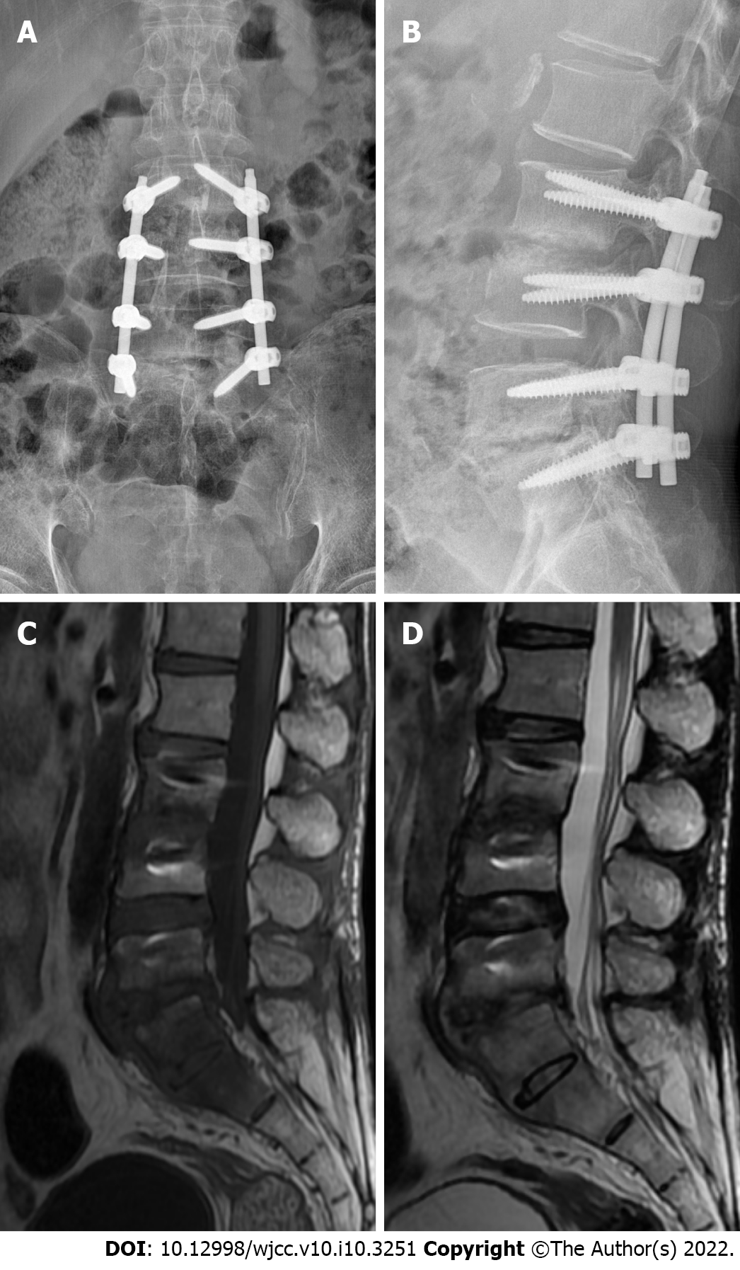
Figure 3 Medical imaging examinations at follow-up.
A and B: X-ray image showing that the position of fixation in the L3–S1 vertebral body was good; C and D: Magnetic resonance imaging showing increased density of the intervertebral spaces of L3/L4 and L5/S1, with a weak T1 signal. No recurrence was noted.
- Citation: Shi XW, Li ST, Lou JP, Xu B, Wang J, Wang X, Liu H, Li SK, Zhen P, Zhang T. Scedosporium apiospermum infection of the lumbar vertebrae: A case report. World J Clin Cases 2022; 10(10): 3251-3260
- URL: https://www.wjgnet.com/2307-8960/full/v10/i10/3251.htm
- DOI: https://dx.doi.org/10.12998/wjcc.v10.i10.3251


