Copyright
©The Author(s) 2022.
World J Clin Cases. Apr 6, 2022; 10(10): 3241-3250
Published online Apr 6, 2022. doi: 10.12998/wjcc.v10.i10.3241
Published online Apr 6, 2022. doi: 10.12998/wjcc.v10.i10.3241
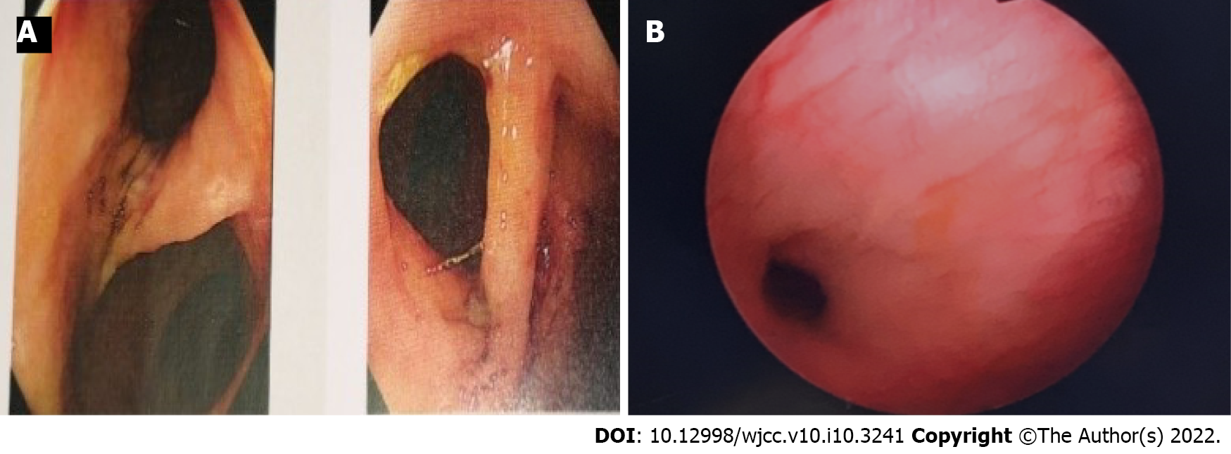
Figure 1 Endoscope.
A: Colonoscopy. A fistula approximately 1.5 cm in size could be seen on the sigmoid colon 20 cm from the anus. The endoscope could pass through the fistula, and the bladder mucosa could be seen. Another fistula with a size of approximately 1.2 cm could be seen 15 cm away from the anus. This suggested multiple colovesical fistulas; B: Cystoscopy. Turbidity of the urine in the bladder, with a large amount of feces visible, and the wall of the bladder was edematous. A fistula could be seen in the lateral wall of the bladder. Both ureteral orifices were fissured, and the release of urine was adequate.
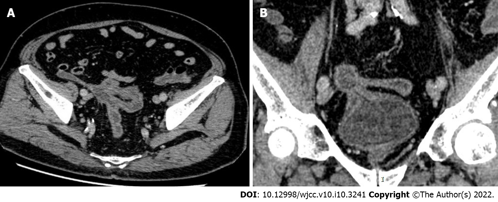
Figure 2 Contrast-enhanced computed tomography of the abdominal pelvis: the appendix was not clearly visualized.
A: The distal sigmoid colon-cecum, right posterior to the top wall of the bladder - sigmoid colon, and middle sigmoid colon-cecal wall were adhered, and fistulas could be seen among these structures. The correlative intestinal wall was thickened, and a contrast-enhanced computed tomography (CT) scan showed enhancement; B: A gas density shadow could be seen in the bladder. The correlative bladder wall was thickened, and a contrast-enhanced CT scan showed enhancement.
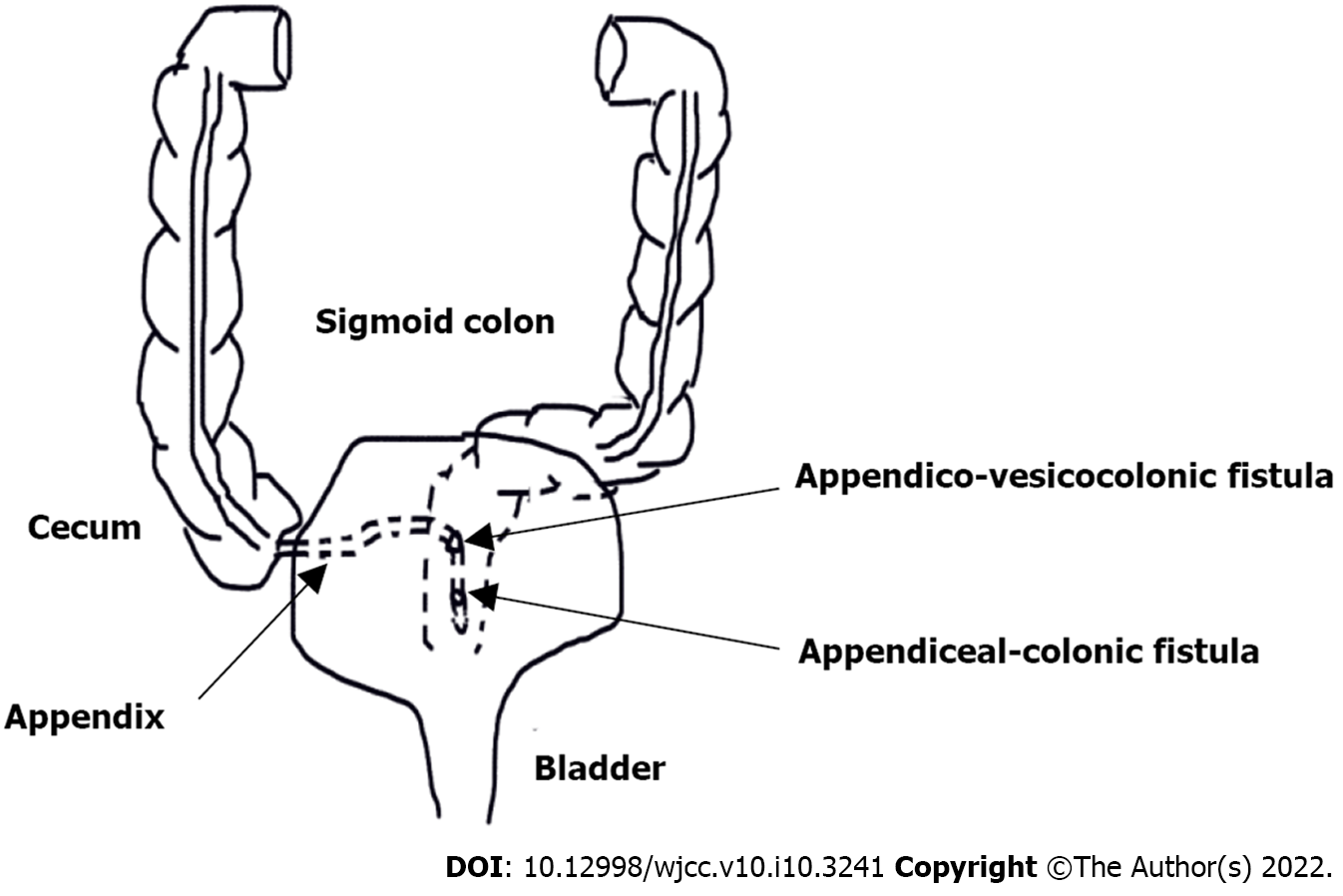
Figure 3 A schematic drawing depicting the fistulous paths between the various organs.
The fistula 20 cm away from the anus was an appendico-vesicocolonic fistula with a diameter of 1.5 cm, and the fistula 15 cm away from the anus was an appendiceal-colonic fistula with a diameter of 1.2 cm.
- Citation: Yan H, Wu YC, Wang X, Liu YC, Zuo S, Wang PY. Appendico-vesicocolonic fistula: A case report and review of literature. World J Clin Cases 2022; 10(10): 3241-3250
- URL: https://www.wjgnet.com/2307-8960/full/v10/i10/3241.htm
- DOI: https://dx.doi.org/10.12998/wjcc.v10.i10.3241


