Copyright
©The Author(s) 2022.
World J Clin Cases. Apr 6, 2022; 10(10): 3213-3221
Published online Apr 6, 2022. doi: 10.12998/wjcc.v10.i10.3213
Published online Apr 6, 2022. doi: 10.12998/wjcc.v10.i10.3213
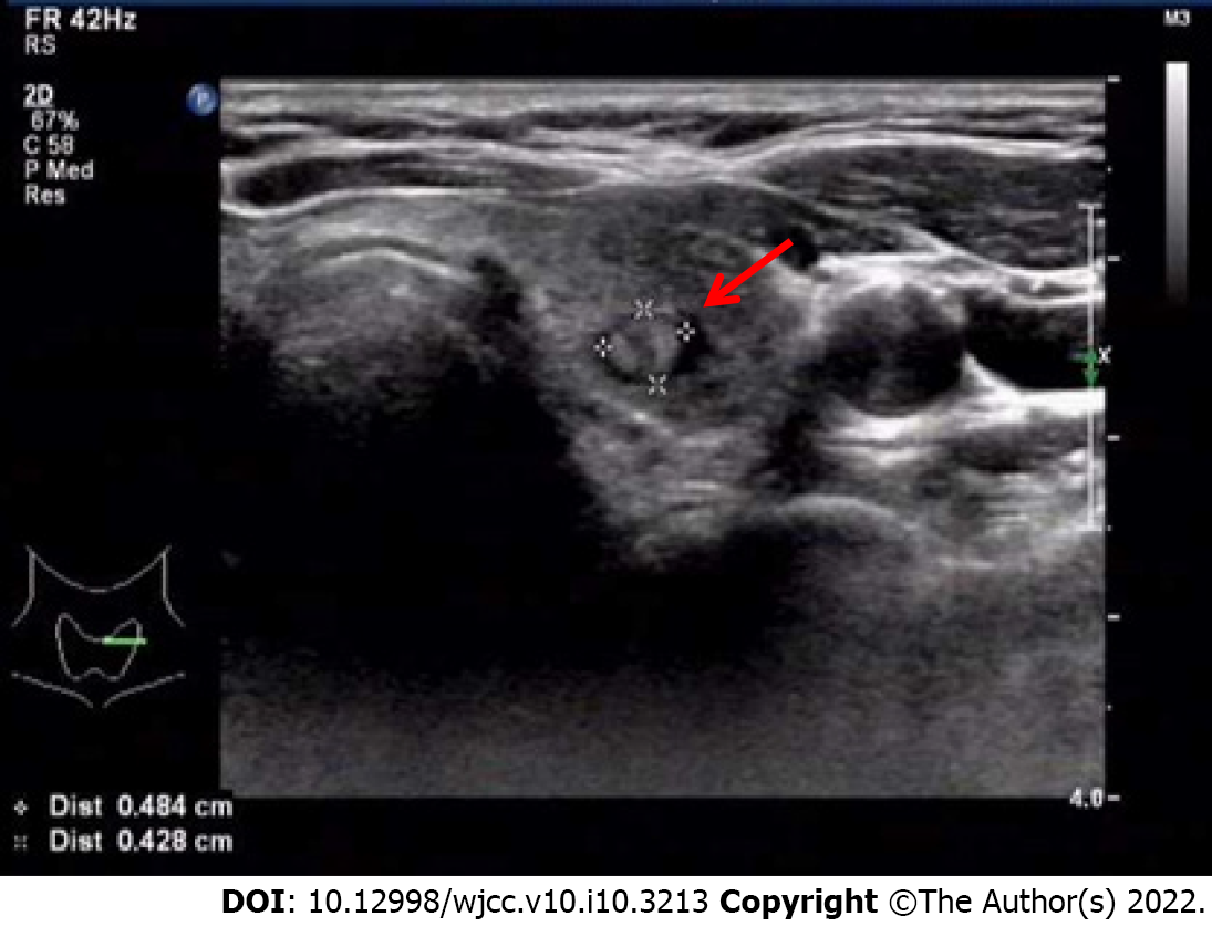
Figure 1 A solid nodule in the left lobe of the thyroid by ultrasound examination.
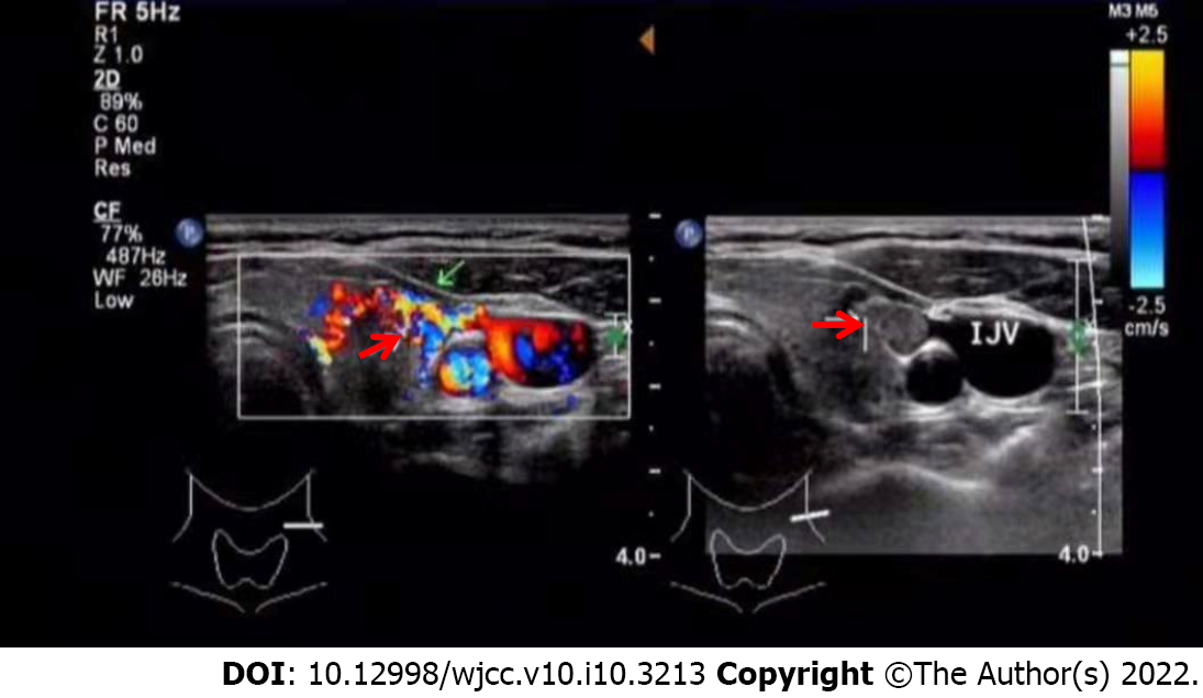
Figure 2 Ultrasound examination revealed a medially echoic mass in the middle thyroid vein.
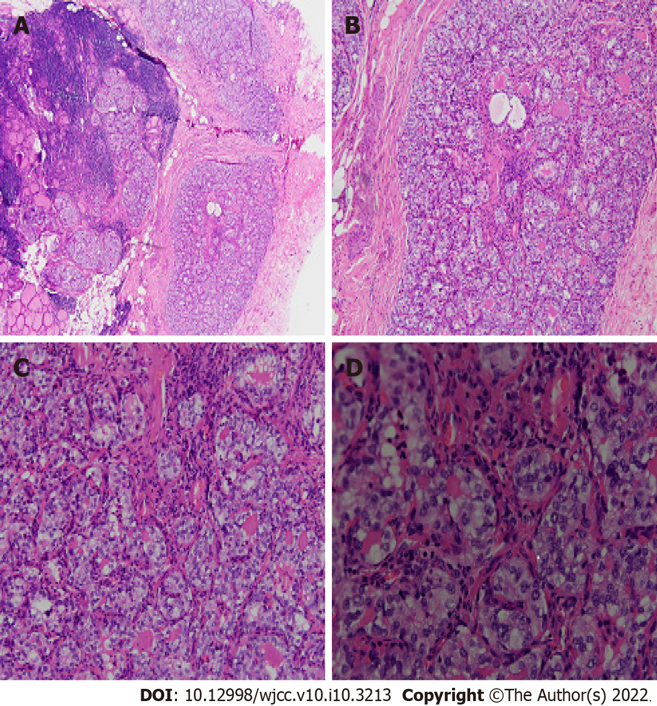
Figure 3 Hematoxylin and eosin staining of left lobe thyroid mass, it shows papillary thyroid microcarcinoma.
A: 4×; B: 10×; C: 20×; D: 40×.
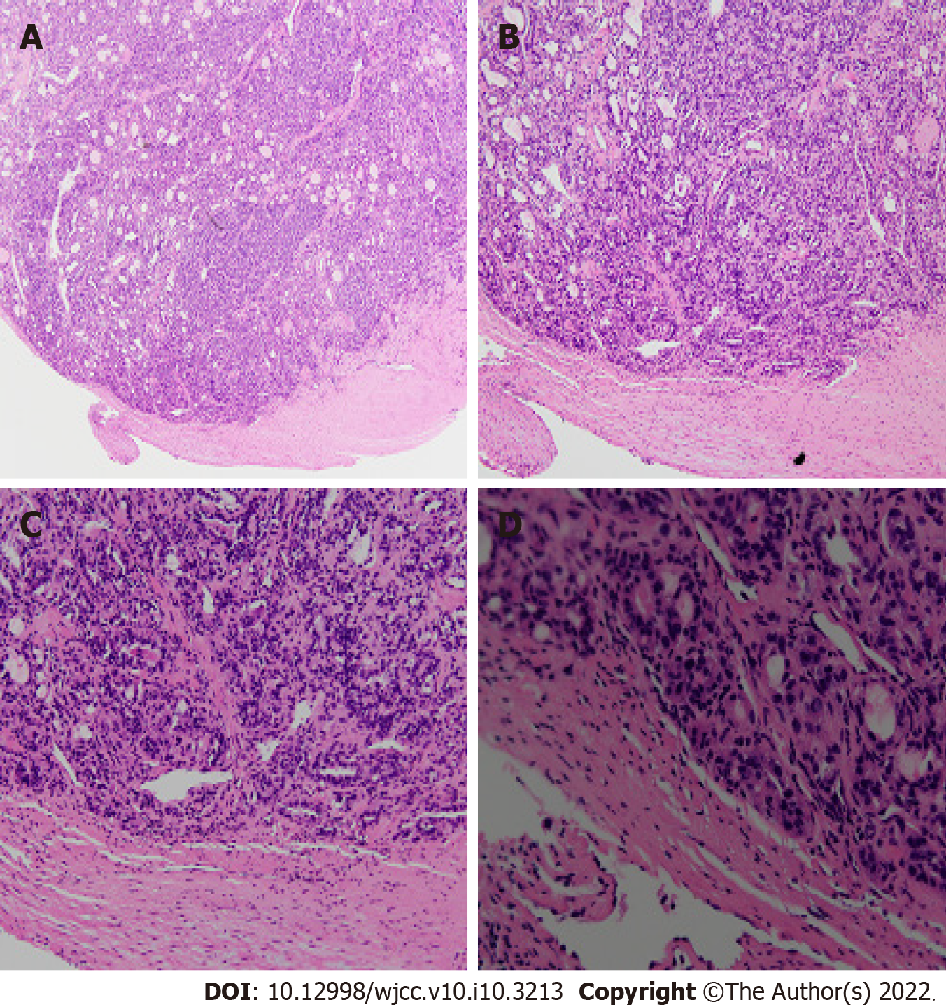
Figure 4 Hematoxylin and eosin staining of the thrombus, it shows carcinoma tissues.
A: 4×; B: 10×; C: 20×; D: 40×.
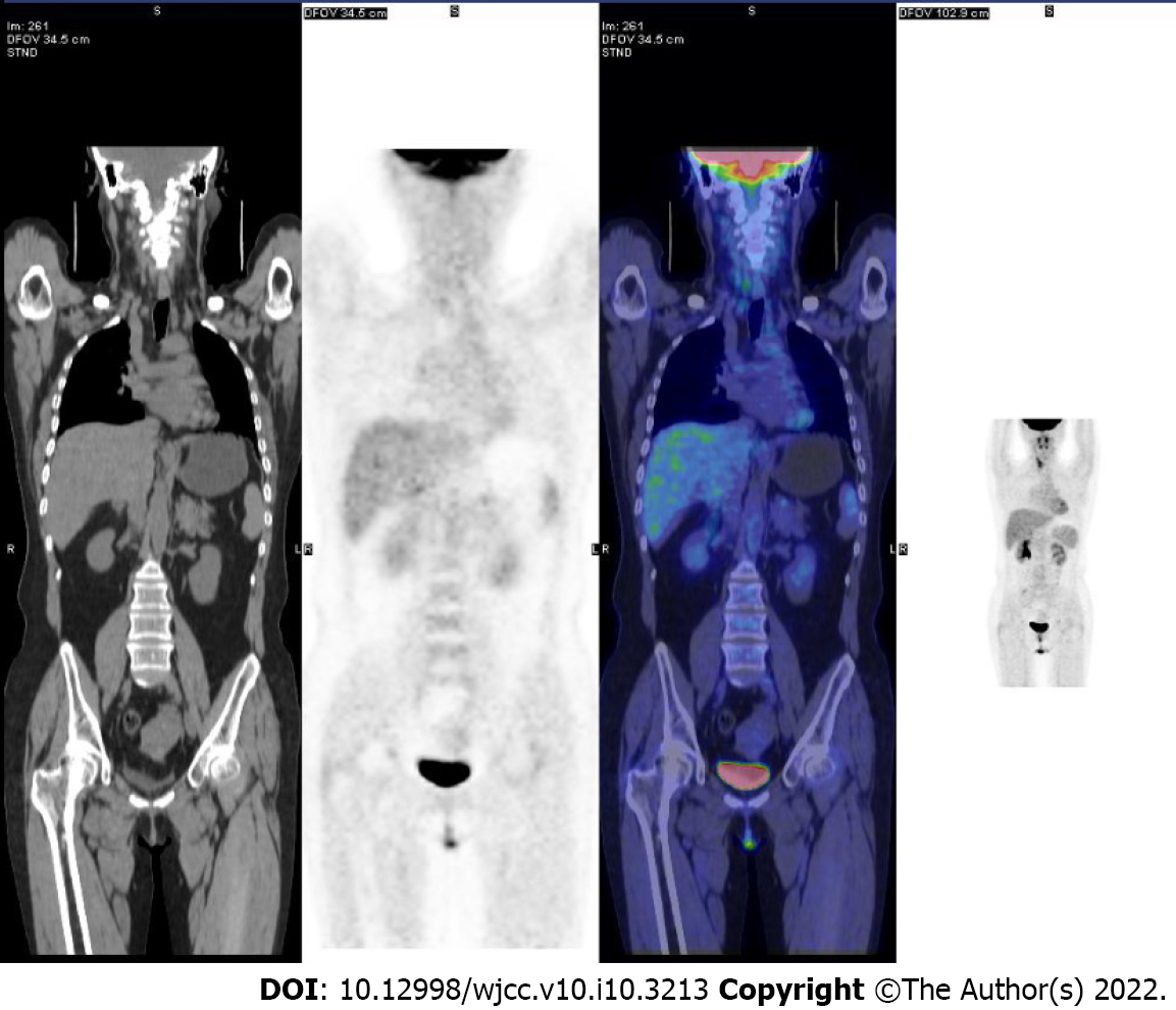
Figure 5 Systemic positron emission tomography metabolism imaging showed no obvious signs of malignancy.
- Citation: Gui Y, Wang JY, Wei XD. Middle thyroid vein tumor thrombus in metastatic papillary thyroid microcarcinoma: A case report and review of literature. World J Clin Cases 2022; 10(10): 3213-3221
- URL: https://www.wjgnet.com/2307-8960/full/v10/i10/3213.htm
- DOI: https://dx.doi.org/10.12998/wjcc.v10.i10.3213


