Copyright
©The Author(s) 2022.
World J Clin Cases. Apr 6, 2022; 10(10): 3200-3205
Published online Apr 6, 2022. doi: 10.12998/wjcc.v10.i10.3200
Published online Apr 6, 2022. doi: 10.12998/wjcc.v10.i10.3200
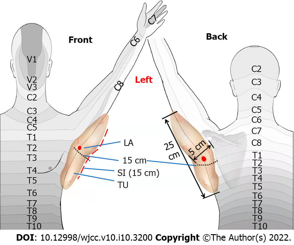
Figure 1 A schematic diagram of tumor dermatomes.
The tumor reached the medial midpoint of forearm, the distribution area of the fifth thoracic vertebral nerve, the lateral edge of scapula and the axillary midline; the tumor size was 25, 15, and 5 cm in length, width and depth, respectively. LA: Local anesthesia (the site of local infiltration anesthesia during surgery); TU: Tumor; SI: Surgical incision.
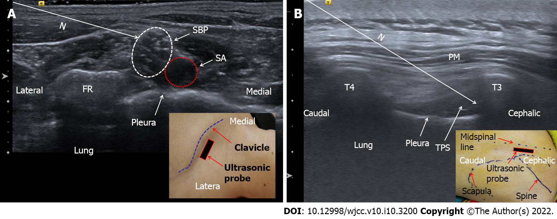
Figure 2 Ultrasound-guided nerve block.
A: Ultrasound-guided supraclavicular brachial plexus block; B: Ultrasound-guided T3-4 paravertebral block. N: Puncture path; SBP: Supraclavicular brachial plexus; SA: Subclavian artery; FR: First rib; PM: Paraspinal muscle; T3: The third thoracic vertebra (transverse process); T4: The fourth thoracic vertebra (transverse process); TPS: Thoracic paravertebral space.
- Citation: Liu Q, Zhong Q, Zhou NN, Ye L. Giant tumor resection under ultrasound-guided nerve block in a patient with severe asthma: A case report. World J Clin Cases 2022; 10(10): 3200-3205
- URL: https://www.wjgnet.com/2307-8960/full/v10/i10/3200.htm
- DOI: https://dx.doi.org/10.12998/wjcc.v10.i10.3200


