Copyright
©The Author(s) 2022.
World J Clin Cases. Apr 6, 2022; 10(10): 3188-3193
Published online Apr 6, 2022. doi: 10.12998/wjcc.v10.i10.3188
Published online Apr 6, 2022. doi: 10.12998/wjcc.v10.i10.3188
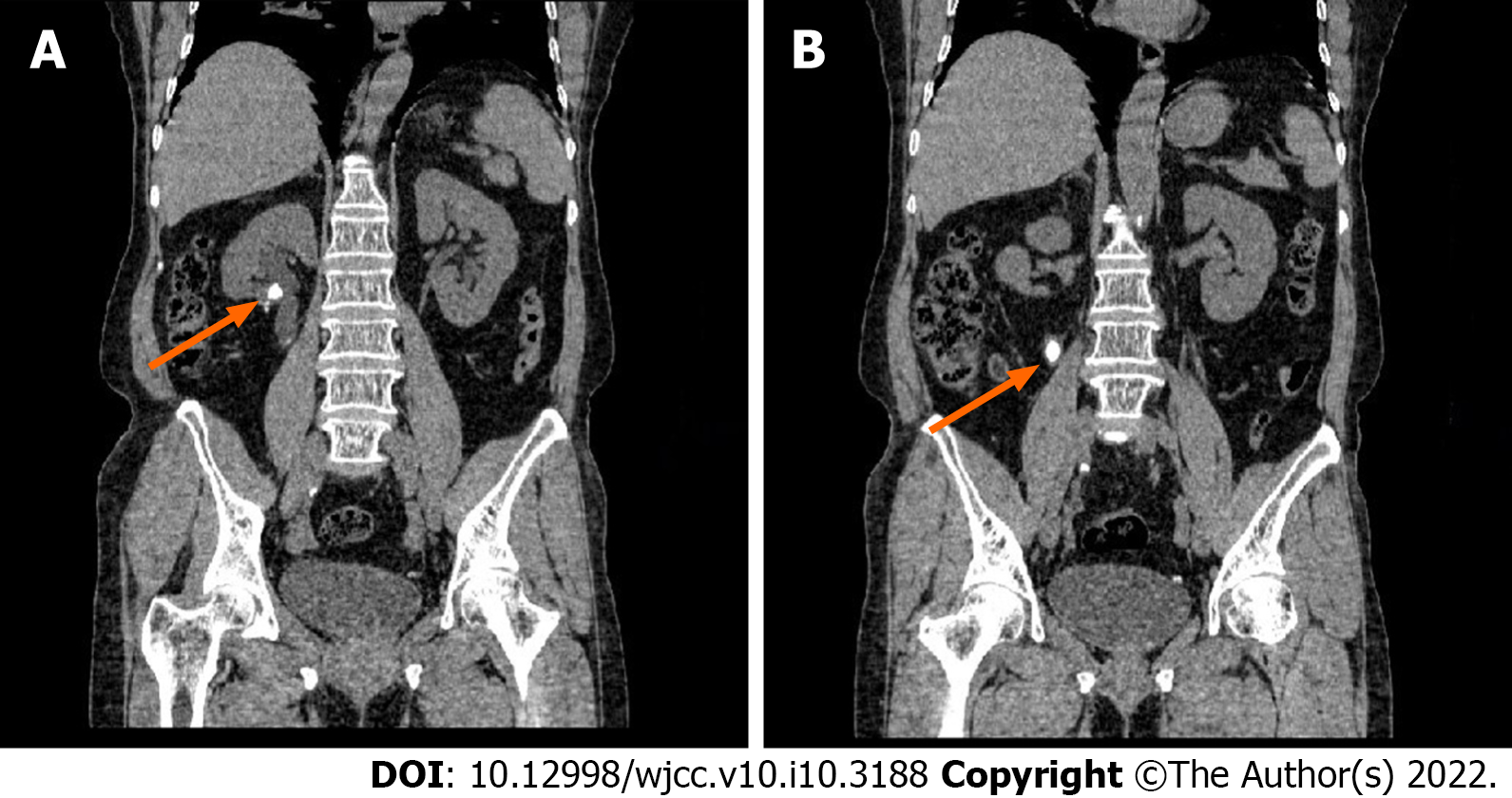
Figure 1 Abdominal computed tomography images demonstrating that the lower pole of the right kidney is absent after operation with a high-density fringe, and two opacities can be seen in the right renal pelvis and proximal ureter (as indicated by arrowheads).
A: 1295 HU; B: 1335 HU.
Figure 2 Intraoperative process of treating calculus.
A: Intraoperative image of percutaneous nephrolithotomy shows that the center of the stones was a white strip funicular substance; B: The foreign body was taken out by use of a forceps; C: It was confirmed as a medium-sized Hem-o-Lok clip via in vitro examination.
- Citation: Sun J, Zhao LW, Wang XL, Huang JG, Fan Y. Migration of a Hem-o-Lok clip to the renal pelvis after laparoscopic partial nephrectomy: A case report. World J Clin Cases 2022; 10(10): 3188-3193
- URL: https://www.wjgnet.com/2307-8960/full/v10/i10/3188.htm
- DOI: https://dx.doi.org/10.12998/wjcc.v10.i10.3188


