Copyright
©The Author(s) 2022.
World J Clin Cases. Apr 6, 2022; 10(10): 3170-3177
Published online Apr 6, 2022. doi: 10.12998/wjcc.v10.i10.3170
Published online Apr 6, 2022. doi: 10.12998/wjcc.v10.i10.3170
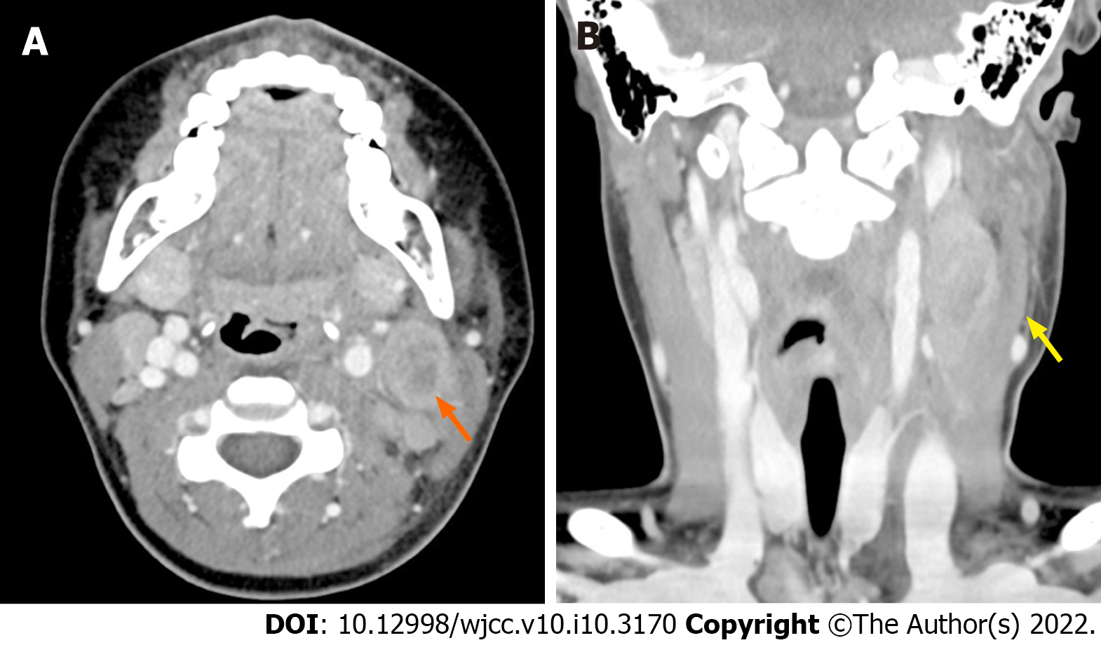
Figure 1 Neck computed tomography horizontal (A) and coronal (B) views showing perilesional infiltration (yellow arrow) and necrosis (orange arrow) as acute infectious lymphadenitis.
Neck computed tomography reveals a 2.5 cm × 2.0 cm × 3.2 cm-sized acute infectious lymphadenitis with perilesional infiltration and necrosis.
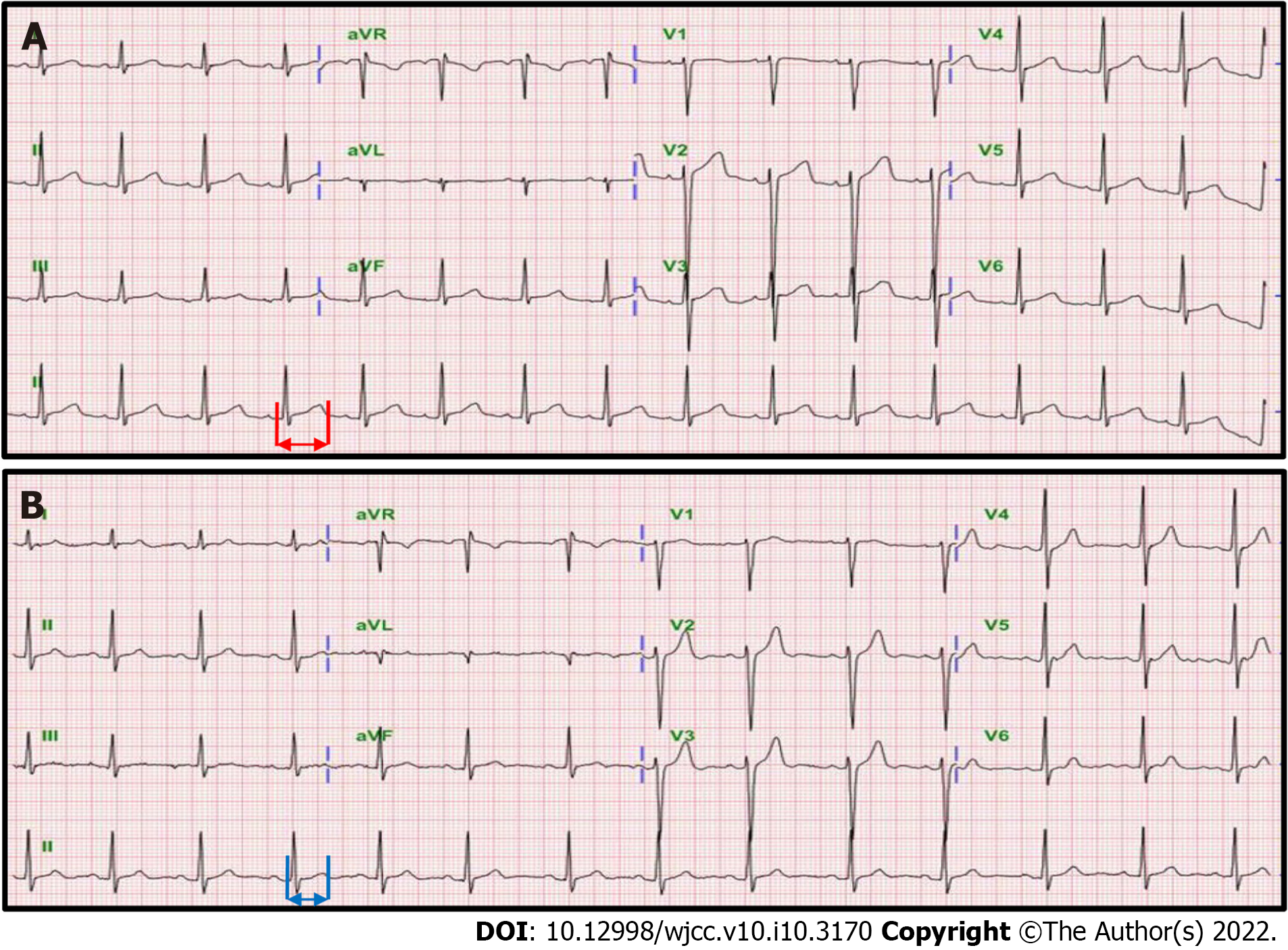
Figure 2 Serial echocardiography findings.
A: Prolonged QT interval was detected on echocardiography on hospital day 4 (red arrow); B: QT interval was gradually shortened in serial echocardiography at 2 month after discharge and finally completely normalized 2 years after discharge (blue arrow).
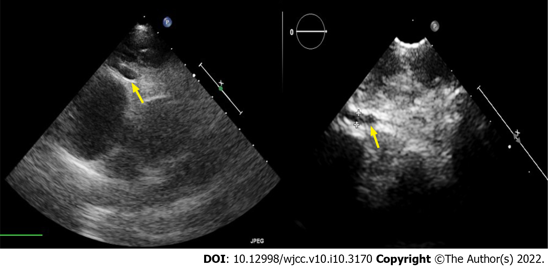
Figure 3 Echocardiography showing right coronary artery ectasia (yellow arrow).
Dilated right coronary artery diameter (3.99 mm-4.73 mm) and left coronary artery diameter (2.35 mm) are observed.
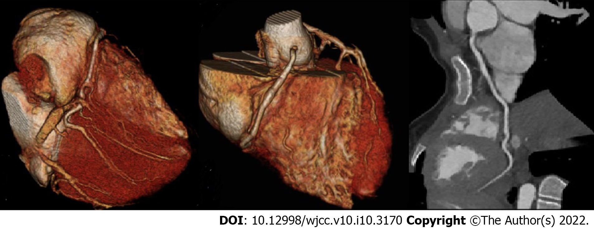
Figure 4 Multifocal ectatic change in the whole right coronary artery (maximum diameter: 3.
5 mm).
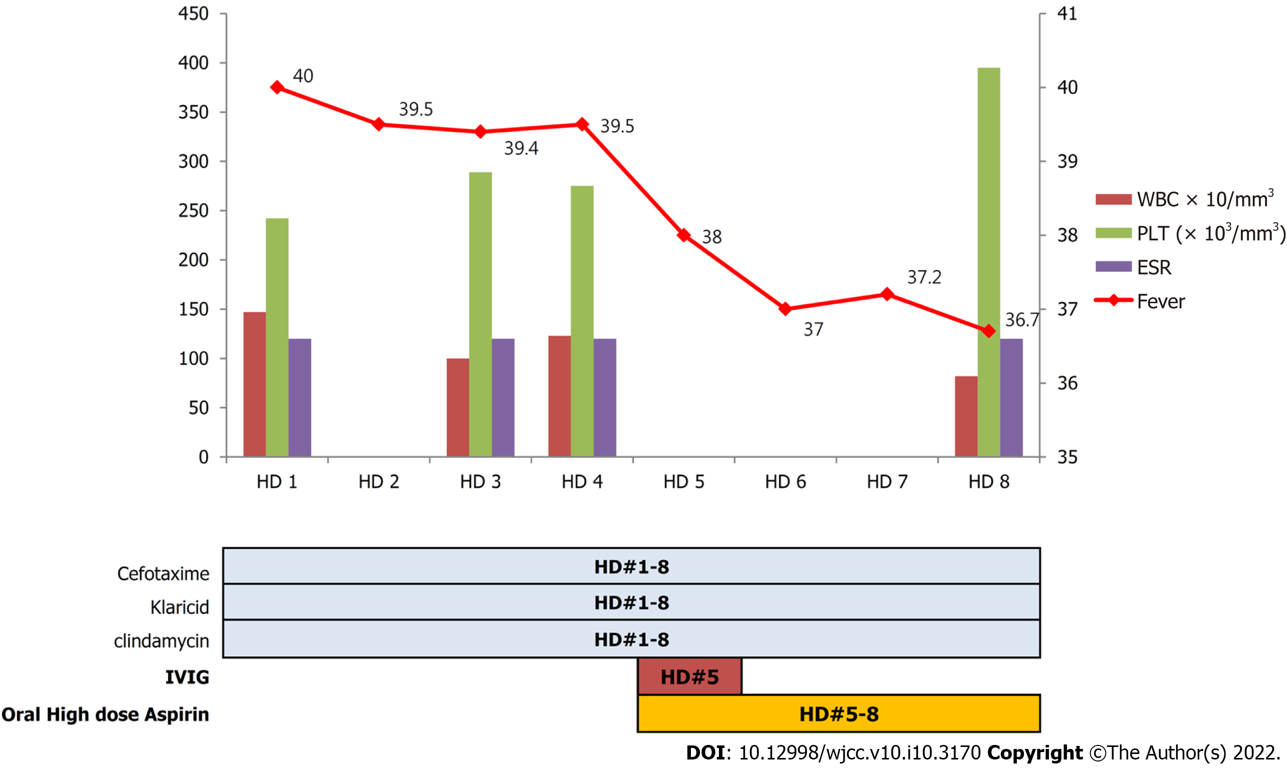
Figure 5 Chronological evolution of the case including symptoms, laboratory data, and treatment during hospitalization.
WBC: White blood cell; PLT: Platelet; ESR: Erythrocyte sedimentation rate; IVIG: Intravenous immunoglobulin.
- Citation: Kim N, Choi YJ, Na JY, Oh JW. Lymph-node-first presentation of Kawasaki disease in a 12-year-old girl with cervical lymphadenitis caused by Mycoplasma pneumoniae: A case report. World J Clin Cases 2022; 10(10): 3170-3177
- URL: https://www.wjgnet.com/2307-8960/full/v10/i10/3170.htm
- DOI: https://dx.doi.org/10.12998/wjcc.v10.i10.3170


